Review
. 2021 Jan:19:157-168.
doi: 10.1016/j.jtos.2020.05.009. Epub 2020 May 26.

The role of Th17 immunity in chronic ocular surface disorders

Affiliations
Review

The role of Th17 immunity in chronic ocular surface disorders

Nai-Wen Fan et al. Ocul Surf. 2021 Jan.

Abstract

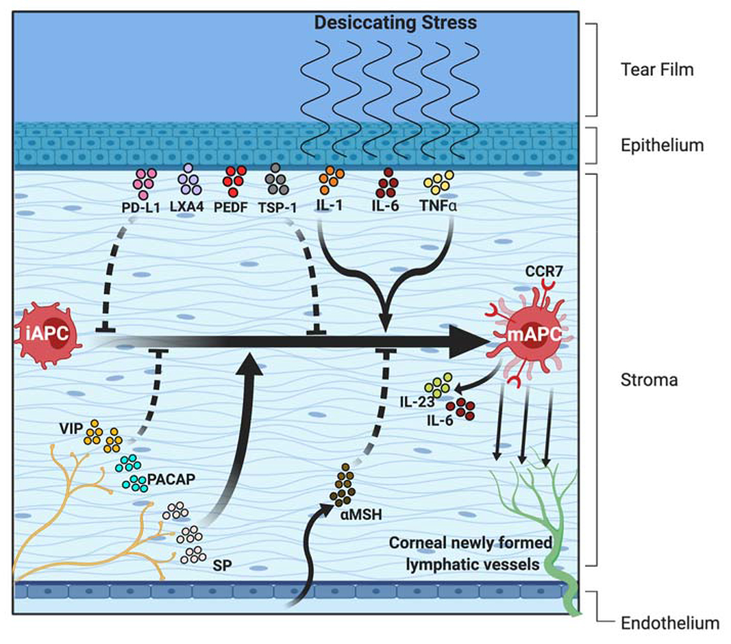
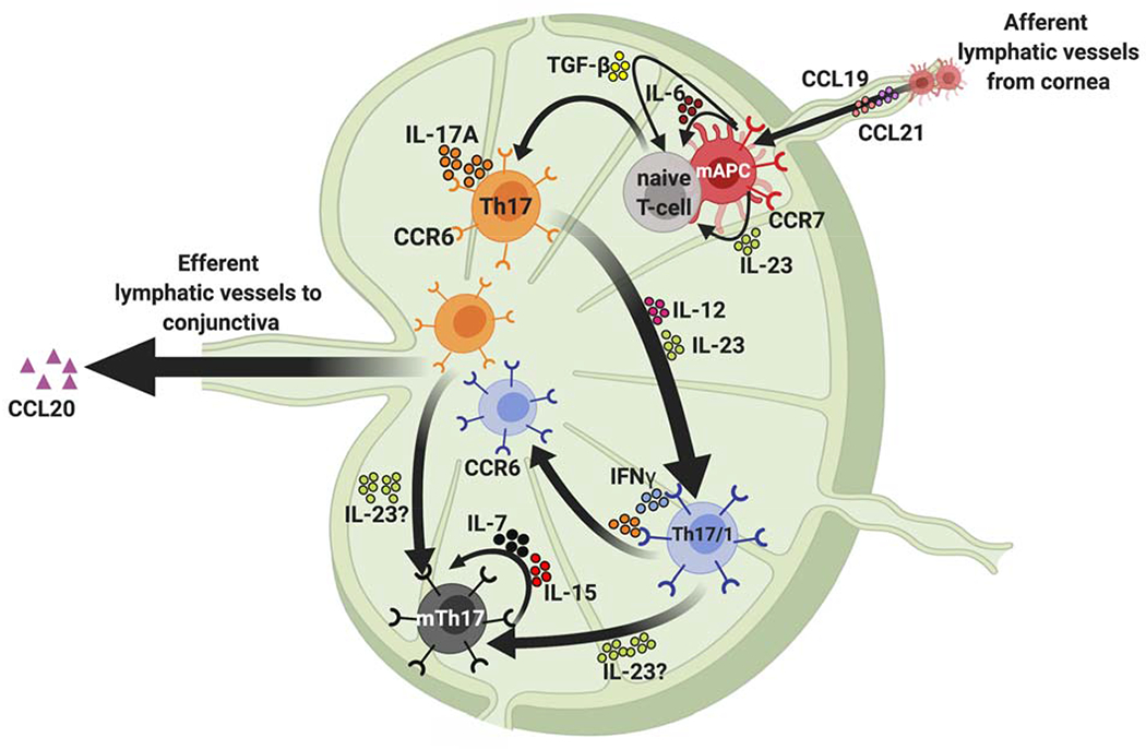
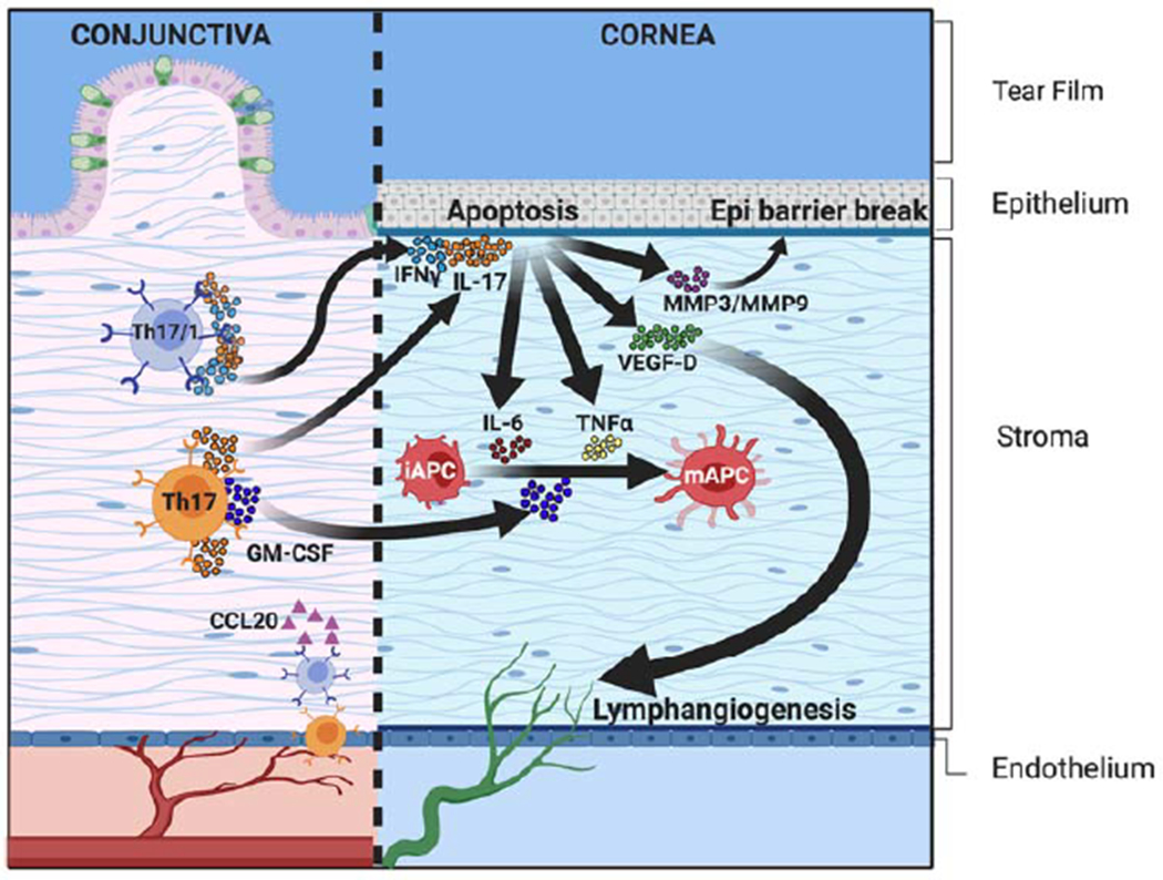
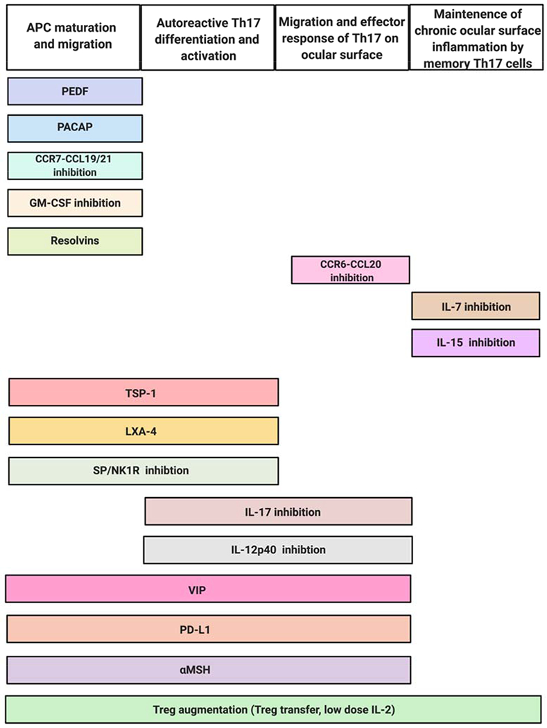
Th17 cells have been implicated in the pathogenesis of numerous inflammatory and autoimmune conditions. At the ocular surface, Th17 cells have been identified as key effector cells in chronic ocular surface disease. Evidence from murine studies indicates that following differentiation and expansion, Th17 cells migrate from the lymphoid tissues to the eye, where they release inflammatory cytokines including, but not limited to, their hallmark cytokine IL-17A. As the acute phase subsides, a population of long-lived memory Th17 cells persist, which predispose hosts both to chronic inflammation and severe exacerbations of disease; of great interest is the small subset of Th17/1 cells that secrete both IL-17A and IFN-γ in acute-on-chronic disease exacerbation. Over the past decade, substantial progress has been made in deciphering how Th17 cells interact with the immune and neuroimmune pathways that mediate chronic ocular surface disease. Here, we review (i) the evidence for Th17 immunity in chronic ocular surface disease, (ii) regulatory mechanisms that constrain the Th17 immune response, and (iii) novel therapeutic strategies targeting Th17 cells.

Keywords: Chronic ocular surface disorders; Dry eye disease; IL-17; Immunological memory; Immunoregulation; Th17.


Conflict of interest statement

Conflicts of interest: Massachusetts Eye and Ear owns intellectual property related to anti-inflammation of targeting IL-17 and substance P in ocular surface diseases. R.D. is consultant to Dompé, Aldeyra, Santen, and holds equity in Claris Biotherapeutics and Aramis Biosciences.

Figures

Figure 1:
Figure 1:. In the afferent arm of the Th17 response, activated APCs are poised to migrate from the ocular surface to the draining lymphoid tissue.
In homeostatic conditions, the cornea contains a heterogenous population of immature antigen presenting cells (iAPCs) with limited capacity to stimulate T cells. However, these cells acquire MHC class II, CD80 and CD86 expression on exposure to inflammatory stress to generate mature APCs (mAPCs). The upregulated expression of MHC class-II and costimulatory molecules has been attributed to IL-1, IL-6, and TNF-α from corneal epithelial cells, Substance P (SP) from the corneal nerves, and granulocyte-macrophage colony-stimulating factor (GM-CSF) from Th17 cells (not shown in the figure). Regarding immunoregulatory factors, various molecules including PD-L1, LXA4, PEDF, and TSP-1 derived from corneal epithelium, VIP and PACAP expressed by corneal nerves, and αMSH from the aqueous humor play an immunosuppressive role by limiting inflammatory cytokine-induced APC maturation. The chemokine receptor CCR7 expressed by mature APCs directs their trafficking to the lymph nodes via newly formed corneal lymphatic vessels.
Figure 2:
Figure 2:. Generation of autoreactive Th17 effector and memory cells.
During Th17 cell development, naïve T cells migrate from the thymus and differentiate into Th17 cells in the draining lymphoid tissue on engagement with activated APCs and exposure to the local inflammatory cytokine milieu. TGF-β, IL-6 and IL-23 expressed by the APCs in the draining lymph node play a critical role in the development of pathogenic Th17 cells. Once generated, the migration of CCR6+ Th17 cells to the ocular surface is facilitated by the attractant chemokine CCL-20 at the ocular surface. In addition, activated Th17 cells produce IFN-γ under the influence of IL-12 and IL-23, and these Th17/1 cells contribute to disease exacerbation. In the chronic stage, effector Th17 cells develop into memory Th17 cells (mTh17), which are generated from both Th17 and Th17/1 subsets and maintained by IL-7 and IL-15. These mTh17 cells mediate the chronic inflammation in DED.
Figure 3:
Figure 3:. Migration and effector response of Th17 cells at the ocular surface.
Th17 and Th17/1 cells express high levels of IL-17A (by Th17 and Th17/1) and IFN-γ (by Th17/1 cells). These inflammatory cytokines mediate the apoptosis of corneal epithelial cells both directly and indirectly (via the proteolytic matrix metalloproteinases [MMP-3 and MMP-9]). IL-17A also contributes to lymphangiogenesis by upregulating the expression of VEGF-D in the cornea, which facilitates the continuous migration of pro-inflammatory cells from the ocular surface to the draining lymphoid tissue. Th17 cells express GM-CSF, which promotes the maturation of iAPCs, and further increases ocular surface inflammation.
Figure 4:
Figure 4:. Novel strategies suppressing Th17 to limit ocular surface inflammation.
Multiple promising therapeutic strategies aim to ameliorate ocular surface inflammation by regulating the differentiation, activation, migration or effector responses of Th17 cells.

Similar articles

Cited by

References

    1. Gipson IK. The ocular surface: the challenge to enable and protect vision: the Friedenwald lecture. Invest Ophthalmol Vis Sci 2007;48:4390; 4391–8. 10.1167/iovs.07-0770. - DOI - PMC - PubMed
    1. Khanna RC. Ocular surface disorders. Community Eye Heal 2017;30:S1–2. - PMC - PubMed
    1. Skalicky SE, Goldberg I, McCluskey P. Ocular surface disease and quality of life in patients with glaucoma. Am J Ophthalmol 2012;153:1–9.e2. 10.1016/j.ajo.2011.05.033. - DOI - PubMed
    1. Baudouin C Detrimental effect of preservatives in eyedrops: implications for the treatment of glaucoma. Acta Ophthalmol 2008;86:716–26. 10.1111/j.1755-3768.2008.01250.x. - DOI - PubMed
    1. Benítez-Del-Castillo J, Labetoulle M, Baudouin C, Rolando M, Akova YA, Aragona P, et al. Visual acuity and quality of life in dry eye disease: Proceedings of the OCEAN group meeting. Ocul Surf 2017;15:169–78. 10.1016/j.jtos.2016.11.003. - DOI - PubMed

Publication types