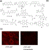
Fluorogenic probes for imaging cellular phosphatase activity
- PMID: 32470893
- PMCID: PMC7483602
- DOI: 10.1016/j.cbpa.2020.04.004
Fluorogenic probes for imaging cellular phosphatase activity
Abstract
The ability to visualize enzyme activity in a cell, tissue, or living organism can greatly enhance our understanding of the biological roles of that enzyme. While many aspects of cellular signaling are controlled by reversible protein phosphorylation, our understanding of the biological roles of the protein phosphatases involved is limited. Here, we provide an overview of progress toward the development of fluorescent probes that can be used to visualize the activity of protein phosphatases. Significant advances include the development of probes with visible and near-infrared (near-IR) excitation and emission profiles, which provides greater tissue and whole-animal imaging capabilities. In addition, the development of peptide-based probes has provided some selectivity for a phosphatase of interest. Key challenges involve the difficulty of achieving sufficient selectivity for an individual member of a phosphatase enzyme family and the necessity of fully validating the best probes before they can be adopted widely.
Keywords: Cellular imaging; Fluorescent probe; Protein phosphatase.
Copyright © 2020 Elsevier Ltd. All rights reserved.
Conflict of interest statement
Conflict of interest statement Nothing declared.
Figures


References
-
- Walsh C, Posttranslational Modification of Proteins: Expanding Nature’s Inventory, first ed., Greenwood Village, Colorado, Roberts and Co. Publishers, 2006.
-
- Barr AJ, Protein tyrosine phosphatases as drug targets: strategies and challenges of inhibitor development, Future Med. Chem 2 (2010) 1563–1576. - PubMed
-
- Xu A-J, Xia X-H, Du S-T, Gu J-C, Clinical significance of PHPT1 protein expression in lung cancer, Chin. Med. J. (Engl.) 123 (2010) 3247–3251. - PubMed
Publication types
MeSH terms
Substances
Grants and funding
LinkOut - more resources
Full Text Sources

