

A Chronic Fetal Leucine Infusion Potentiates Fetal Insulin Secretion and Increases Pancreatic Islet Size, Vascularity, and β Cells in Late-Gestation Sheep
- PMID: 32470982
- PMCID: PMC7398779
- DOI: 10.1093/jn/nxaa138
A Chronic Fetal Leucine Infusion Potentiates Fetal Insulin Secretion and Increases Pancreatic Islet Size, Vascularity, and β Cells in Late-Gestation Sheep
Abstract
Background: Infusion of a complete amino acid mixture into normal late-gestation fetal sheep potentiates glucose-stimulated insulin secretion (GSIS). Leucine acutely stimulates insulin secretion in late-gestation fetal sheep and isolated fetal sheep islets in vitro.
Objectives: We hypothesized that a 9-d leucine infusion would potentiate GSIS in fetal sheep.
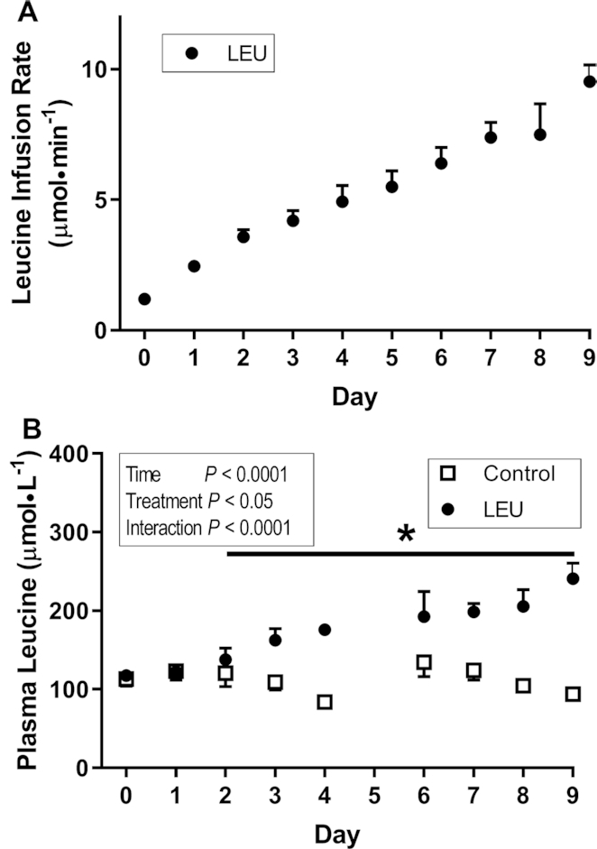
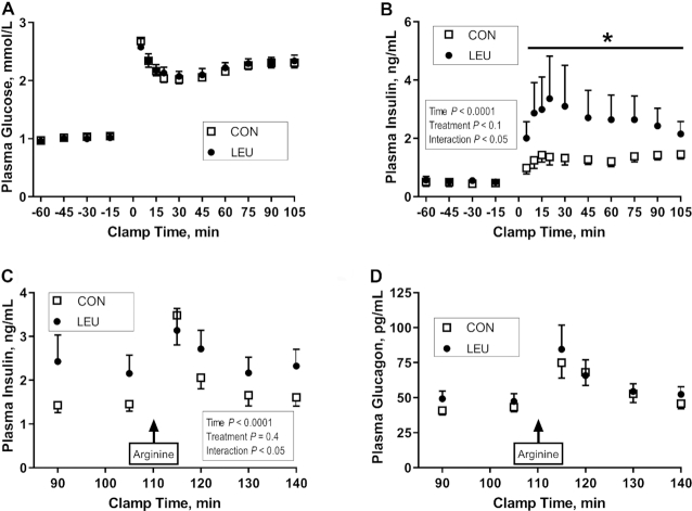
Methods: Columbia-Rambouillet fetal sheep at 126 days of gestation received a 9-d leucine infusion to achieve a 50%-100% increase in leucine concentrations or a control infusion. At the end of the infusion we measured GSIS, pancreatic morphology, and expression of pancreatic mRNAs. Pancreatic islet endothelial cells (ECs) were isolated from fetal sheep and incubated with supplemental leucine or vascular endothelial growth factor A (VEGFA) followed by collection of mRNA. Data measured at multiple time points were compared with a repeated-measures 2-factor ANOVA. Data measured at 1 time point were compared using Student's t test or the Mann-Whitney test.
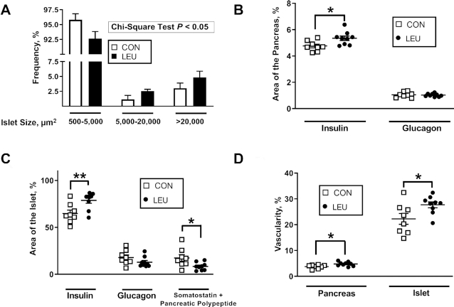
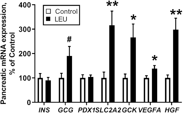
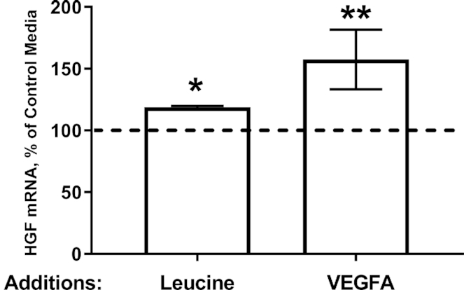
Results: Glucose-stimulated insulin concentrations were 80% higher in leucine-infused (LEU) fetuses than in controls (P < 0.05). In the pancreas, LEU fetuses had a higher proportion of islets >5000 μm2 than controls (75% more islets >5000 μm2; P < 0.05) and a larger proportion of the pancreas that stained for β cells (12% greater; P < 0.05). Pancreatic and pancreatic islet vascularity were both 25% greater in LEU fetuses (P < 0.05). Pancreatic VEGFA and hepatocyte growth factor (HGF) mRNA expressions were 38% and 200% greater in LEU fetuses than in controls (P < 0.05), respectively. In isolated islet ECs, HGF mRNA was 20% and 50% higher after incubation in supplemental leucine (P < 0.05) or VEGFA (P < 0.01), respectively.
Conclusions: A 9-d leucine infusion potentiates fetal GSIS, demonstrating that glucose and leucine act synergistically to stimulate insulin secretion in fetal sheep. A greater proportion of the pancreas being comprised of β cells and higher pancreatic vascularity contributed to the higher GSIS.
Keywords: VEGFA; fetus; hepatocyte growth factor; insulin; islet; leucine; pancreas; pregnancy; vascularity; β cell.
Copyright © The Author(s) on behalf of the American Society for Nutrition 2020.
Figures
References
-
- Fowden AL. Effects of adrenaline and amino acids on the release of insulin in the sheep fetus. J Endocrinol. 1980;87:113–21. - PubMed
-
- Gresores A, Anderson S, Hood D, Zerbe GO, Hay WW Jr. Separate and joint effects of arginine and glucose on ovine fetal insulin secretion. Am J Physiol. 1997;272:E68–73. - PubMed
-
- Philipps AF, Dubin JW, Raye JR. Alanine-stimulated insulin secretion in the fetal and neonatal lamb. Am J Obstet Gynecol. 1980;136:597–602. - PubMed
-
- Rozance PJ, Limesand SW, Hay WW Jr. Decreased nutrient-stimulated insulin secretion in chronically hypoglycemic late-gestation fetal sheep is due to an intrinsic islet defect. Am J Physiol Endocrinol Metab. 2006;291:E404–11. - PubMed
