

Magnetic Resonance Imaging and Modeling of the Glymphatic System
- PMID: 32471025
- PMCID: PMC7344900
- DOI: 10.3390/diagnostics10060344
Magnetic Resonance Imaging and Modeling of the Glymphatic System
Abstract
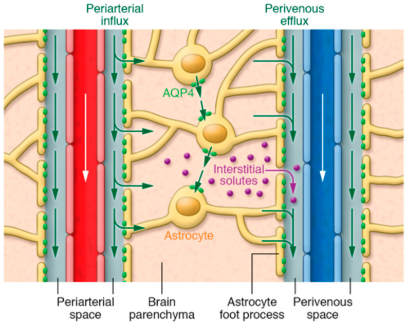
The glymphatic system is a newly discovered waste drainage pathway in the brain; it plays an important role in many neurological diseases. Ongoing research utilizing various cerebrospinal fluid tracer infusions, either directly or indirectly into the brain parenchyma, is investigating clearance pathways by using distinct imaging techniques. In the present review, we discuss the role of the glymphatic system in various neurological diseases and efflux pathways of brain waste clearance based on current evidence and controversies. We mainly focus on new magnetic resonance imaging (MRI) modeling techniques, along with traditional computational modeling, for a better understanding of the glymphatic system function. Future sophisticated modeling techniques hold the potential to generate quantitative maps for glymphatic system parameters that could contribute to the diagnosis, monitoring, and prognosis of neurological diseases. The non-invasive nature of MRI may provide a safe and effective way to translate glymphatic system measurements from bench-to-bedside.
Keywords: Keywords: glymphatic system; aquaporin-4; bulk flow; cerebrospinal fluid; computational modeling; contrast-enhanced MRI; diffusion; extracellular space; flow pathways; neurological diseases.
Conflict of interest statement
The authors declare no conflict of interest. The funders had no role in the design of the study; in the collection, analyses, or interpretation of data; in the writing of the manuscript, or in the decision to publish the results.
Figures
Similar articles
-
MRI and glymphatic system.Stroke Vasc Neurol. 2019 Apr 5;4(2):75-77. doi: 10.1136/svn-2018-000197. eCollection 2019 Jul. Stroke Vasc Neurol. 2019. PMID: 31338214 Free PMC article. Review.
-
Glymphatic MRI in idiopathic normal pressure hydrocephalus.Brain. 2017 Oct 1;140(10):2691-2705. doi: 10.1093/brain/awx191. Brain. 2017. PMID: 28969373 Free PMC article.
-
The Association between Glymphatic System and Perivascular Macrophages in Brain Waste Clearance.Diagnostics (Basel). 2024 Mar 29;14(7):731. doi: 10.3390/diagnostics14070731. Diagnostics (Basel). 2024. PMID: 38611644 Free PMC article.
-
Monitoring the perivascular cerebrospinal fluid dynamics of the glymphatic pathway using co-localized photoacoustic microscopy.Opt Lett. 2023 May 1;48(9):2265-2268. doi: 10.1364/OL.486129. Opt Lett. 2023. PMID: 37126250
-
Glymphatic imaging using MRI.J Magn Reson Imaging. 2020 Jan;51(1):11-24. doi: 10.1002/jmri.26892. Epub 2019 Aug 18. J Magn Reson Imaging. 2020. PMID: 31423710 Review.
Cited by
-
A window into the brain: multimodal MRI assessment of vascular cognitive impairment.Front Neurosci. 2025 Apr 16;19:1526897. doi: 10.3389/fnins.2025.1526897. eCollection 2025. Front Neurosci. 2025. PMID: 40309660 Free PMC article. Review.
-
Waste Clearance in the Brain.Front Neuroanat. 2021 Jul 7;15:665803. doi: 10.3389/fnana.2021.665803. eCollection 2021. Front Neuroanat. 2021. PMID: 34305538 Free PMC article. Review.
-
The meningeal lymphatic vessels and the glymphatic system: Potential therapeutic targets in neurological disorders.J Cereb Blood Flow Metab. 2022 Aug;42(8):1364-1382. doi: 10.1177/0271678X221098145. Epub 2022 Apr 28. J Cereb Blood Flow Metab. 2022. PMID: 35484910 Free PMC article. Review.
-
Effect of Various Lengths of Respiration on Heart Rate Variability during Simple Bhramari (Humming).Int J Yoga. 2023 May-Aug;16(2):123-131. doi: 10.4103/ijoy.ijoy_113_23. Epub 2023 Nov 21. Int J Yoga. 2023. PMID: 38204770 Free PMC article.
-
Therapeutic non-invasive brain treatments in Alzheimer's disease: recent advances and challenges.Inflamm Regen. 2022 Oct 3;42(1):31. doi: 10.1186/s41232-022-00216-8. Inflamm Regen. 2022. PMID: 36184623 Free PMC article. Review.
References
-
- Bakker E.N., Bacskai B.J., Arbel-Ornath M., Aldea R., Bedussi B., Morris A.W., Weller R.O., Carare R.O. Lymphatic Clearance of the Brain: Perivascular, Paravascular and Significance for Neurodegenerative Diseases. Cell Mol. Neurobiol. 2016;36:181–194. doi: 10.1007/s10571-015-0273-8. - DOI - PMC - PubMed
-
- Javed K., Lui F. Neuroanatomy, Choroid Plexus. StatPearls; Treasure Island, FL, USA: 2020. - PubMed
-
- Iliff J.J., Wang M., Liao Y., Plogg B.A., Peng W., Gundersen G.A., Benveniste H., Vates G.E., Deane R., Goldman S.A., et al. A paravascular pathway facilitates CSF flow through the brain parenchyma and the clearance of interstitial solutes, including amyloid beta. Sci. Transl. Med. 2012;4:147ra111. doi: 10.1126/scitranslmed.3003748. - DOI - PMC - PubMed
Publication types
Grants and funding
LinkOut - more resources
Full Text Sources
