. 2020 May 27;12(6):1371.
doi: 10.3390/cancers12061371.

Progression-Dependent Altered Metabolism in Osteosarcoma Resulting in Different Nutrient Source Dependencies

Affiliations

Progression-Dependent Altered Metabolism in Osteosarcoma Resulting in Different Nutrient Source Dependencies

Raphaela Fritsche-Guenther et al. Cancers (Basel). .

Abstract

Osteosarcoma (OS) is a primary malignant bone tumor and OS metastases are mostly found in the lung. The limited understanding of the biology of metastatic processes in OS limits the ability for effective treatment. Alterations to the metabolome and its transformation during metastasis aids the understanding of the mechanism and provides information on treatment and prognosis. The current study intended to identify metabolic alterations during OS progression by using a targeted gas chromatography mass spectrometry approach. Using a female OS cell line model, malignant and metastatic cells increased their energy metabolism compared to benign OS cells. The metastatic cell line showed a faster metabolic flux compared to the malignant cell line, leading to reduced metabolite pools. However, inhibiting both glycolysis and glutaminolysis resulted in a reduced proliferation. In contrast, malignant but non-metastatic OS cells showed a resistance to glycolytic inhibition but a strong dependency on glutamine as an energy source. Our in vivo metabolic approach hinted at a potential sex-dependent metabolic alteration in OS patients with lung metastases (LM), although this will require validation with larger sample sizes. In line with the in vitro results, we found that female LM patients showed a decreased central carbon metabolism compared to metastases from male patients.

Keywords: GC-MS; flux analysis; glucose; glutamine, sex and gender; osteosarcoma.


Conflict of interest statement

The authors declare no conflict of interest.

Figures
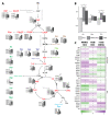
Figure 1
Figure 1
Enhanced metabolism in malignant and metastatic OS cells. (A) Levels of annotated metabolites of glycolysis, tricarboxylic acid (TCA) cycle, glycerol and amino acids in human osteosarcoma HOS (H), N-methyl-N’-nitro-N-nitrosoguanidine (MNNG)/HOS (M) and 143B (1) cells (each comprising n = 3 biological and n = 2 technical replicates). Data shows the mean and standard deviation of the normalized peak areas. Samples were analyzed using an unpaired Student’s t-test, with a p ≤ 0.05 deemed statistically significant and indicated with a star (*). (B) Number of metabolites after unit scaling of normalized peak areas (in total and according to the subsets of metabolic pathways comprising glycolysis (glyc), TCA and amino acids (AA). Metabolites with higher peak areas than the average for that metabolite are shown above zero and those with lower peak areas below. We used the Pearson’s chi-squared test to perform a chi-squared contingency table test and goodness-of-fit with a threshold of p < 0.05 for statistically relevant differences between the metabolic groups and the total number of metabolites (indicated with a star (*)). Y-axis: frequency of metabolites above or below individual metabolite mean average. (C) Heat map from proteins associated with the central carbon metabolism comparing MNNG/HOS to HOS, 143B to HOS and 143B to MNNG/HOS (MNNG) cells (each n = 5 replicates). Ratios from the log2 values of the label-free intensities from the biological replicates are shown. Samples were analyzed using an unpaired Student’s t-test with a p < 0.05 deemed statistically significant and indicated with a star (*). 2HG: 2-hydroxy-glutaric acid. 2PGA: glyceric-acid-2-phosphate. 3PGA: glyceric-acid-3-phosphate. 13BPG: 1/3-bis-phosphoglyceric acid. aCoA: acetyl-CoenzymeA. ACO: aconitase. Ala: alanine. ALDO: aldolase. aKG: alpha-ketoglutaric acid. Asn: asparagine. Asp: aspartic acid. Cit: citric acid. CS: citrate synthase. DHAP: dihydroxyacetonephosphate. DLST: dihydrolipoamide succinyltransferase (E2) component of the 2-oxoglutarate dehydrogenase complex. ENO: enolase. F6P: fructose-6-phosphate. F16BP: fructose-1/6-bisphopsphate. FH: fumarate hydratase. Fum: fumaric acid. G6P: glucose-6-phosphate. GA3P: glyceraldehyde-3-phosphate. GAPDH: glycerinaldehyde-3-phosphate dehydrogenase. Glc: glucose. Gln: glutamine. Glu: glutamic acid. Glyc3P: glycerol-3-phosphate. Gly: glycine. Glyc: glycerol. GPI: glucose-6-phosphate isomerase. HK: hexokinase. IDH: isocitrate dehydrogenase. Ile: isoleucine. Lac: lactic acid. Leu: leucine. Lys: lysine. Mal: malic acid. Met: methionine. OAA: oxaloacetate. OGDH: 2-oxoglutarate dehydrogenase (E1) component of the 2-oxoglutarate dehydrogenase complex.Phe: phenylalanine. PEP: phosphoenol-pyruvic acid. PFK: phosphofructokinase. PGK: phosphoglyceratkinase. PGM: phosphoglycerate mutase. PKM: pyruvate kinase. Pyr: pyruvic acid. Pro: proline. SDH: succinate dehydrogenase. Ser: serine. Suc: succinic acid. SUCL: succinyl-CoA synthetase. TCA: tricarboxylic acid. Thr: threonine. TPI: triosephosphatisomerase. Tyr: tyrosine. Val: valine. R5P: ribose-5-phosphate. Rib5P: ribulose-5-phosphate.
Figure 2
Figure 2
Enhanced lipid metabolism in malignant and metastatic osteosarcoma (OS) cells. Measured compounds comparing MNNG/HOS to HOS, 143B to HOS and 143B to the MNNG/HOS cells. For each cell type, n = 5 replicates were measured. Data are log2 values from the ratios calculated from the mean of the normalized areas (liquid chromatography measurement) or intensities (flow injection analysis measurement). AA: amino acids. BA: biogenic amines. DG: diglycerides. LPC: lysophosphatidylcholines. PC: phosphatidylcholines. SM: sphingomyelins. TG: triglycerides.
Figure 3
Figure 3
Increased metabolic flux cycle in metastatic OS cells. (A,E,F) Label incorporation of glycolytic intermediates in OS cells in the presence of 13C-glucose for 30 min (A) or 13C-glutamine for 60 min (E,F). Each n = 3 replicates. (B) Log2 label-free intensities were used to calculate the ratio of MNNG/HOS (M) to HOS (H), 143B (1) to HOS and 143B to the MNNG/HOS cells. Each n = 5 replicates. (C,D) Mean fluorescence intensity (MFI) of phosphorylated mechanistic target of rapamycin (P-mTOR) and phosphorylated extracellular signal related kinases 1 and 2 (P-ERK1/2). Each n = 3 replicates. Samples were analyzed using an unpaired Student’s t-test with a p ≤ 0.05 deemed statistically significant (indicated by a star (*)). 2-HG: 2-hydroxy-glutaric acid. Ala: alanine. aKG: alpha-ketoglutaric acid. Cit: citric acid. Fum: fumaric acid. G6P: glucose-6-phosphate. Lac: lactic acid. Mal: malic acid. PHGDH: D-3-phosphoglycerate dehydrogenase. PSAT1: phosphoserine aminotransferase. Pyr: pyruvic acid. R5P: ribulose-5-phosphate. Ser: serine. Suc: succinic acid.
Figure 4
Figure 4
Progression-dependent switch in nutrient sources. (A) Viability of osteosarcoma cell lines HOS, MNNG/HOS and 143B after glucose (glc) or glutamine (gln) starvation for 48 h. For each condition, n = 4 replicates were measured. (B) Cells were treated with 0.25 mM 3-Bromopyruvic acid (BrPy) or control solvent phosphate buffered saline. For each condition, n = 3 replicates were measured. Samples were analyzed using an unpaired Student’s t-test, with a p ≤ 0.05 deemed statistically significant (indicated by the star (*)). OD: optical density.
Figure 5
Figure 5
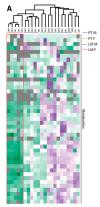
Sex-dependent metabolic alteration in OS patients with primary tumors (PT) and lung metastases (LM). (A) Heat map for the unsupervised hierarchical clustering of the n = 7 PT and n = 5 LM (males = M and females = F). Perseus version 1.4.0.20 was used for clustering. The Z-score from the normalized peak areas from the technical and biological replicates for the annotated metabolites from the central carbon metabolism was calculated. Negative Z-scores were displayed in green, positive Z-Scores in purple. An unweighted average linkage clustering and Euclidean distance preprocessed with k-means was used for the hierarchical clustering. (B) Levels of annotated metabolites of glycolysis, the TCA cycle, glycerol and amino acids in n = 7 PT and n = 5 LM from males and females. Data show the mean and standard deviation of the normalized peak area. Samples were analyzed using an unpaired Student’s t-test, with a p ≤ 0.05 deemed as statistically significant and indicated by the star (*). 2HG: 2-hydroxy-glutaric acid. 2PGA: glyceric-acid-2-phosphate. 3PGA: glyceric-acid-3-phosphate. 13BPG: 1/3-bis-phosphoglyceric acid. aCoA: acetyl-CoA. Ala: alanine. aKG: alpha-ketoglutaric acid. Asn: asparagine. Asp: aspartic acid. Cit: citric acid. Cys: cysteine. DHAP: dihydroxyacetonephosphate. F6P: fructose-6-phosphate. F16BP: fructose-1/6-bisphopsphate. Fum: fumaric acid. G6P: glucose-6-phosphate. GA3P: glyceraldehyde-3-phosphate. Glc: glucose. Gln: glutamine. Glu: glutamic acid. Glyc3P: glycerol-3-phosphate. Gly: glycine. Glyc: glycerol. Ile: isoleucine. Lac: lactic acid. Leu: leucine. Lys: lysine. Mal: malic acid. Met: methionine. OAA: oxaloacetate. Phe: phenylalanine. PEP: phosphoenol-pyruvic acid. Pyr: pyruvic acid. Pro: proline. Ser: serine. Suc: succinic acid. TCA: tricarboxylic acid. Thr: threonine. Trp: tryptophan. Tyr: tyrosine. Val: valine.
Figure 5
Figure 5
Sex-dependent metabolic alteration in OS patients with primary tumors (PT) and lung metastases (LM). (A) Heat map for the unsupervised hierarchical clustering of the n = 7 PT and n = 5 LM (males = M and females = F). Perseus version 1.4.0.20 was used for clustering. The Z-score from the normalized peak areas from the technical and biological replicates for the annotated metabolites from the central carbon metabolism was calculated. Negative Z-scores were displayed in green, positive Z-Scores in purple. An unweighted average linkage clustering and Euclidean distance preprocessed with k-means was used for the hierarchical clustering. (B) Levels of annotated metabolites of glycolysis, the TCA cycle, glycerol and amino acids in n = 7 PT and n = 5 LM from males and females. Data show the mean and standard deviation of the normalized peak area. Samples were analyzed using an unpaired Student’s t-test, with a p ≤ 0.05 deemed as statistically significant and indicated by the star (*). 2HG: 2-hydroxy-glutaric acid. 2PGA: glyceric-acid-2-phosphate. 3PGA: glyceric-acid-3-phosphate. 13BPG: 1/3-bis-phosphoglyceric acid. aCoA: acetyl-CoA. Ala: alanine. aKG: alpha-ketoglutaric acid. Asn: asparagine. Asp: aspartic acid. Cit: citric acid. Cys: cysteine. DHAP: dihydroxyacetonephosphate. F6P: fructose-6-phosphate. F16BP: fructose-1/6-bisphopsphate. Fum: fumaric acid. G6P: glucose-6-phosphate. GA3P: glyceraldehyde-3-phosphate. Glc: glucose. Gln: glutamine. Glu: glutamic acid. Glyc3P: glycerol-3-phosphate. Gly: glycine. Glyc: glycerol. Ile: isoleucine. Lac: lactic acid. Leu: leucine. Lys: lysine. Mal: malic acid. Met: methionine. OAA: oxaloacetate. Phe: phenylalanine. PEP: phosphoenol-pyruvic acid. Pyr: pyruvic acid. Pro: proline. Ser: serine. Suc: succinic acid. TCA: tricarboxylic acid. Thr: threonine. Trp: tryptophan. Tyr: tyrosine. Val: valine.

Similar articles

Cited by

References

    1. Zamborsky R., Kokavec M., Harsanyi S., Danisovic L. Identification of Prognostic and Predictive Osteosarcoma Biomarkers. Med. Sci. 2019;7:28. doi: 10.3390/medsci7020028. - DOI - PMC - PubMed
    1. Kansara M., Teng M.W., Smyth M.J., Thomas D.M. Translational biology of osteosarcoma. Nat. Rev. Cancer. 2014;14:722–735. doi: 10.1038/nrc3838. - DOI - PubMed
    1. Dean D.C., Shen S., Hornicek F.J., Duan Z. From genomics to metabolomics: Emerging metastatic biomarkers in osteosarcoma. Cancer Metastasis Rev. 2018 doi: 10.1007/s10555-018-9763-8. - DOI - PubMed
    1. Saraf A.J., Fenger J.M., Roberts R.D. Osteosarcoma: Accelerating Progress Makes for a Hopeful Future. Front. Oncol. 2018;8:4. doi: 10.3389/fonc.2018.00004. - DOI - PMC - PubMed
    1. Misaghi A., Goldin A., Awad M., Kulidjian A.A. Osteosarcoma: A comprehensive review. Sicot J. 2018;4:12. doi: 10.1051/sicotj/2017028. - DOI - PMC - PubMed

LinkOut - more resources