. 2020 May 27;12(6):1374.
doi: 10.3390/cancers12061374.

Vav1 is Essential for HIF-1α Activation via a Lysosomal VEGFR1-Mediated Degradation Mechanism in Endothelial Cells

Affiliations

Vav1 is Essential for HIF-1α Activation via a Lysosomal VEGFR1-Mediated Degradation Mechanism in Endothelial Cells

Jaewoo Hong et al. Cancers (Basel). .

Abstract

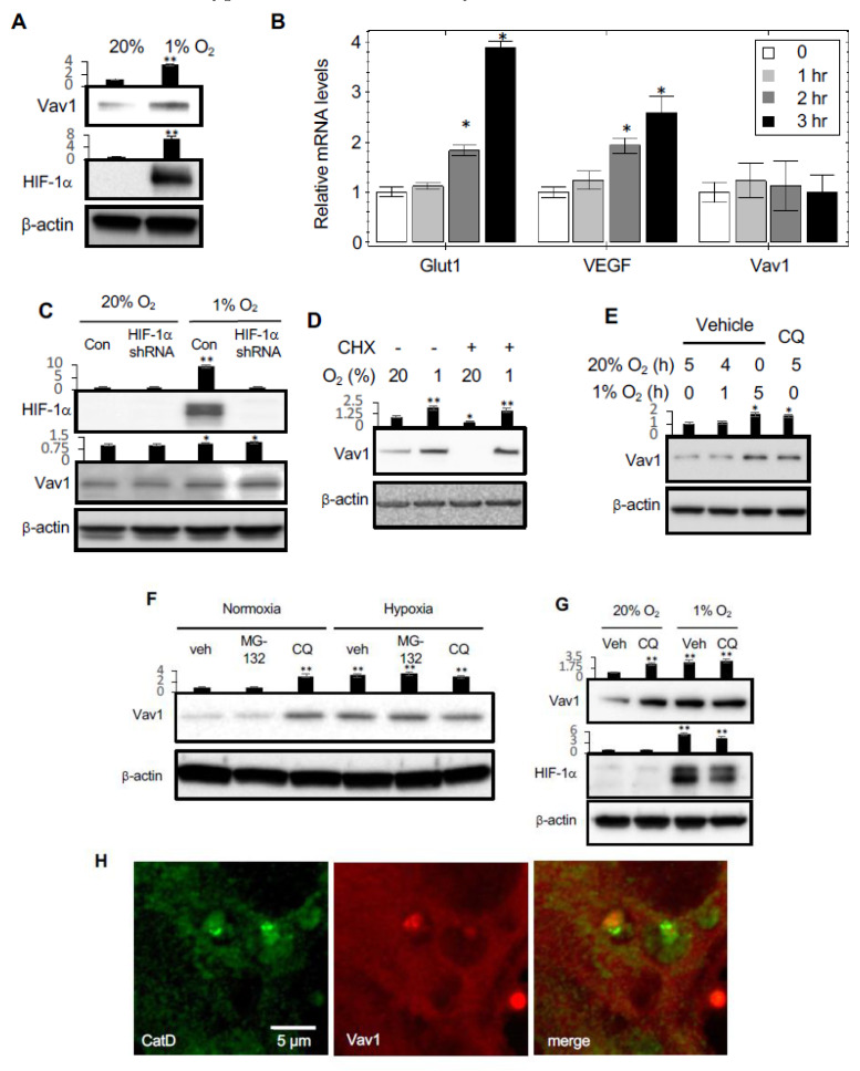
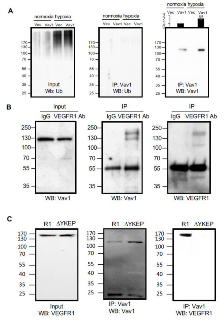
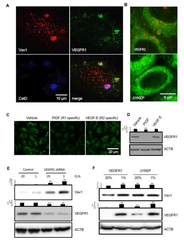
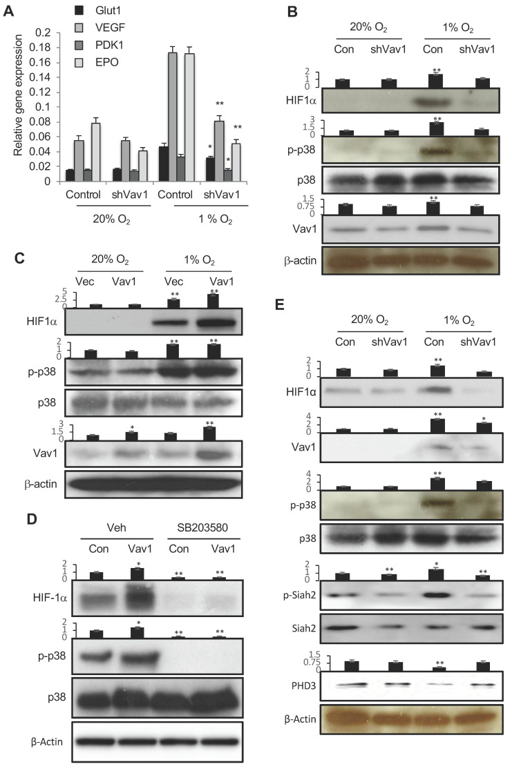
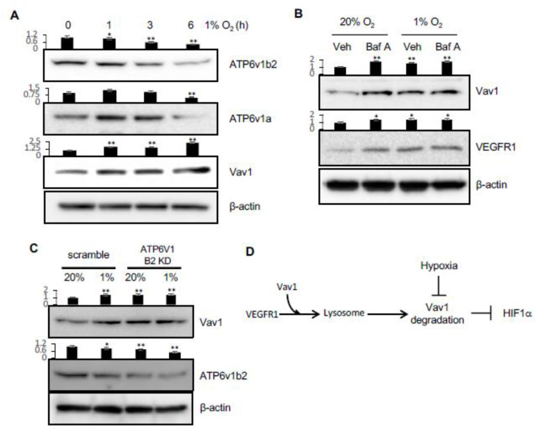
The vascular response to hypoxia and ischemia is essential for maintaining homeostasis during stressful conditions and is particularly critical for vital organs such as the heart. Hypoxia-inducible factor-1 (HIF-1) is a central regulator of the response to hypoxia by activating transcription of numerous target genes, including vascular endothelial growth factor (VEGF). Here we identify the guanine nucleotide exchange factor (GEF) Vav1, a regulator of the small Rho-GTPase and cell signaling in endothelial cells, as a key vascular regulator of hypoxia. We show that Vav1 is present in the vascular endothelium and is essential for HIF-1 activation under hypoxia. So, we hypothesized that Vav1 could be a key regulator of HIF-1 signaling. In our findings, Vav1 regulates HIF-1α stabilization through the p38/Siah2/PHD3 pathway. In normoxia, Vav1 binds to vascular endothelial growth factor receptor 1 (VEGFR1), which directs Vav1 to lysosomes for degradation. In contrast, hypoxia upregulates Vav1 protein levels by inhibiting lysosomal degradation, which is analogous to HIF-1α regulation by hypoxia: both proteins are constitutively produced and degraded in normoxia allowing for a rapid response when stress occurs. Consequently, hypoxia rapidly stabilizes Vav1, which is required for HIF-1α accumulation. This shows that Vav1 is the key mediator controlling the stabilization of HIF1α in hypoxic conditions. With this finding, we report a novel pathway to stabilize HIF-1, which shows a possible reason why clinical trials targeting HIF-1 has not been effective. Targeting Vav1 can be the new approach to overcome hypoxic tumors.

Keywords: HIF-1α; VEGFR; Vav1; endothelial cell; hypoxia; protein degradation.


Conflict of interest statement

The authors declare no conflict of interest.

Figures

Figure 1
Figure 1
Hypoxia upregulates Vav1 through inhibition of lysosome-mediated protein degradation. (A) Human umbilical vein endothelial cells (HUVEC)s were cultured either in 20% or 1% O2 for 5 h, followed by Western blot analysis for the indicated proteins. (B) qPCR analysis of Glut1, VEGF and Vav1 in HUVECs cultured in 1% O2 for 0, 1, 2, or 3 h. * p < 0.05 compared to corresponding time 0 in each group (mean ± SD). (C) The levels of Vav1 and HIF-1α were measured by Western blot of HUVECs transduced with HIF-1α or scramble shRNA expressing Lentivirus for 24 h. (D) HUVECs were incubated in normoxic and hypoxic environments for 5 h in the presence or absence of 10 μM cycloheximide. Protein levels were measured by Western blot from the total lysate. (E) HUVECs were incubated in 20% O2 for 5 h, incubated in 20% O2 for an hour and then moved to 1% O2 for 4 h, incubated in 1% O2 for 5 h. The level of Vav1 in the lysate was compared to HUVECs incubated in 20% O2 in the presence of chloroquine (CQ) at 50 μM for 5 h in order to identify whether Vav1 is affected by lysosomal inhibition. (F) HUVEC were incubated in normoxic or hypoxic conditions for 4 h in the presence of either vehicle control, 5 μM of MG-132, or 50 μM of chloroquine (CQ). The total lysate was subjected to Western blotting to measure Vav1 protein levels. (G) Western blot analysis of Vav1 levels in HUVECs cultured in either 20% or 1% O2 in the absence or presence of Bafilomycin A (Baf A) at 100 nM for 5 h. (H) Immunofluorescent staining for Vav1 (red) and Cathepsin D (green) in HUVECs were imaged by an LSM780 confocal microscope. Each experiment was repeated at least ten times and representative images are shown. Mean ± SD, * p < 0.05, ** p < 0.01. The whole western blot images please find in Figure S1.
Figure 2
Figure 2
Vav1 binds to vascular endothelial growth factor receptor 1 (VEGFR1). (A) Either Flag-tagged Vav1 or empty vector control were transduced into HUVEC cells and incubated under normoxic or hypoxic conditions. Cell lysates were subjected to Vav1 immunoprecipitation and subsequent Western blot analysis for ubiquitin (middle panel) or Vav1 as a control (right panel). Ubiquitin levels were also detected in both control and Vav1 cells exposed to hypoxia from the input of the immunoprecipitation (left panel). (B) VEGFR1 and Vav1 were immunoprecipitated by anti-VEGFR1 antibody or control IgG from the cell lysate of HUVEC cells. Vav1 was probed from the input lysate (left) and pulldown product (center). VEGFR1 was detected from the pulldown product (right). (C) Overexpressed wild type (R1) or YKEP motif deleted (ΔYKEP) VEGFR1 were co-IPed with Vav1 antibody. The input was probed with VEGFR1 antibody (left). The pulldown product was probed against Vav1 (center) or VEGFR1 antibody (right). Mean ± SD, * p < 0.05, ** p < 0.01
Figure 3
Figure 3
VEGFR1 carries Vav1 to lysosomes for degradation. (A) HUVECs were immunostained for Cathepsin D, Vav1 and VEGFR1, and analyzed by confocal microscopy. (B) HeLa cells were co-transfected with expression vectors for Vav1 and wild type VEGFR1 or the Vav1 binding domain deleted construct, ΔYKEP. Cells were stained with Lamp2 (red) and Vav1 (green). (C) HUVECs were stimulated with either PlGF or VEGF-E at 50 ng/mL for 5 h, followed by staining with antibodies for Vav1 (green) and imaged under confocal microscopy. (D) HUVECs were stimulated with either PlGF or VEGF-E at 50 ng/mL for 5 h, followed by Western blot to detect VEGFR1 levels. (E) HUVECs were infected with lentiviral vectors carrying scrambled or VEGFR1 shRNAs and cultured in either 20% O2 or 1% O2 incubators. Cell lysates were analyzed by Western blot for Vav1 and VEGFR1. (F) HUVECs were transfected with expression vectors for Vav1 with either VEGFR1 or ΔYKEP VEGFR1. The levels of Vav1 and VEGFR1 were assessed by Western blot. Mean ± SD, ** p < 0.01. The whole western blot images please find in Figure S2.
Figure 4
Figure 4
Vav1 regulates HIF1α via p38 MAPK. (A) HUVECs were transfected with a control vector or shVav1 vector for 24 h, followed by incubation either in normoxia or hypoxia for another 24 h. HIF-1 target gene expression was analyzed by qRT-PCR. * p < 0.05 and ** p < 0.01 compared to corresponding control transfected cells in hypoxia (mean ± SD). Each experiment was performed in triplicate and repeated three times. (B) Control vector- or shVav1 vector-transfected HUVECs were incubated under normoxia or hypoxia conditions for 24 h. The levels of phosphor-p38, HIF-1α, p38 and Vav1 were analyzed by Western blot. (C) Identical procedures and measurements as in (B) except the cells were transfected with a control vector or Vav1 expression vector. (D) Control or Vav1 expression vector-transfected HUVECs were incubated in the absence or presence of SB203580 at 10 μM in hypoxia for 24 h. The levels of HIF-1α, p38 and phospho-p38 were analyzed by Western blot. (E) Control vector- or shVav1 vector-transfected HUVECs were incubated under normoxic or hypoxic conditions for 24 h. The levels of phospho HIF-1α, Vav1, P-p38, p38, P-siah2, Siah2 and PHD3 were analyzed by Western blot. Mean ± SD, * p < 0.05, ** p < 0.01. The whole western blot images please find in Figure S3.
Figure 5
Figure 5
Hypoxia regulates lysosomal activity via v-ATPase and Vav1 levels. (A) HUVEC cells were incubated in hypoxia for 0, 1, 3 or 6 hours. Vav1, ATP6v1a, and ATP6v1b2 were measured in the total lysates. (B) HUVEC were incubated in normoxia or hypoxia for 5 h in the presence or absence of 100 nM of Bafilomycin A (Baf A). Vav1 and VEGFR1 were measured by Western blot of the total lysates. (C) HUVEC were transduced with ATP6v1b2 shRNA-expressing lentivirus or scrambled shRNA virus for 72 h, and then incubated in normoxia or hypoxia for 5 h. Vav1 and ATP6v1b2 levels were measured by Western blot of the total lysate. (D) VEGFR1 carries Vav1 to the lysosome in the cell. Vav1 is degraded in the lysosome, which downregulates HIF1α. Hypoxia blocks the lysosomal degradation. Mean ± SD, * p < 0.05, ** p < 0.01. The whole western blot images please find in Figure S4.

Similar articles

Cited by

References

    1. Lee S., Chen T.T., Barber C.L., Jordan M.C., Murdock J., Desai S., Ferrara N., Nagy A., Roos K.P., Iruela-Arispe M.L. Autocrine VEGF signaling is required for vascular homeostasis. Cell. 2007;130:691–703. doi: 10.1016/j.cell.2007.06.054. - DOI - PMC - PubMed
    1. Tang N., Wang L., Esko J., Giordano F.J., Huang Y., Gerber H.P., Ferrara N., Johnson R.S. Loss of HIF-1alpha in endothelial cells disrupts a hypoxia-driven VEGF autocrine loop necessary for tumorigenesis. Cancer Cell. 2004;6:485–495. doi: 10.1016/j.ccr.2004.09.026. - DOI - PubMed
    1. Ferrara N., Gerber H.P., LeCouter J. The biology of VEGF and its receptors. Nat. Med. 2003;9:669–676. doi: 10.1038/nm0603-669. - DOI - PubMed
    1. Shibuya M. Vascular endothelial growth factor and its receptor system: Physiological functions in angiogenesis and pathological roles in various diseases. J. Biochem. 2013;153:13–19. doi: 10.1093/jb/mvs136. - DOI - PMC - PubMed
    1. Wu F.T., Stefanini M.O., Mac Gabhann F., Kontos C.D., Annex B.H., Popel A.S. A systems biology perspective on sVEGFR1: Its biological function, pathogenic role and therapeutic use. J. Cell Mol. Med. 2010;14:528–552. doi: 10.1111/j.1582-4934.2009.00941.x. - DOI - PMC - PubMed

LinkOut - more resources