Review
. 2020 May 27;21(11):3797.
doi: 10.3390/ijms21113797.

Alteration of Electron Acceptor Preferences in the Oxidative Half-Reaction of Flavin-Dependent Oxidases and Dehydrogenases

Affiliations
Review

Alteration of Electron Acceptor Preferences in the Oxidative Half-Reaction of Flavin-Dependent Oxidases and Dehydrogenases

Kentaro Hiraka et al. Int J Mol Sci. .

Abstract

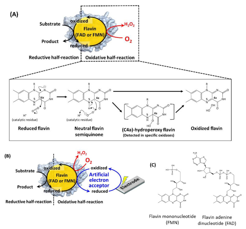
In this review, recent progress in the engineering of the oxidative half-reaction of flavin-dependent oxidases and dehydrogenases is discussed, considering their current and future applications in bioelectrochemical studies, such as for the development of biosensors and biofuel cells. There have been two approaches in the studies of oxidative half-reaction: engineering of the oxidative half-reaction with oxygen, and engineering of the preference for artificial electron acceptors. The challenges for engineering oxidative half-reactions with oxygen are further categorized into the following approaches: (1) mutation to the putative residues that compose the cavity where oxygen may be located, (2) investigation of the vicinities where the reaction with oxygen may take place, and (3) investigation of possible oxygen access routes to the isoalloxazine ring. Among these approaches, introducing a mutation at the oxygen access route to the isoalloxazine ring represents the most versatile and effective strategy. Studies to engineer the preference of artificial electron acceptors are categorized into three different approaches: (1) engineering of the charge at the residues around the substrate entrance, (2) engineering of a cavity in the vicinity of flavin, and (3) decreasing the glycosylation degree of enzymes. Among these approaches, altering the charge in the vicinity where the electron acceptor may be accessed will be most relevant.

Keywords: bioelectrochemistry; dehydrogenase; electron acceptor; enzyme engineering; flavin adenine dinucleotide; flavin mononucleotide; oxidase; oxidative half-reaction; oxygen; oxygen accessible pathway.


Conflict of interest statement

The authors declare no conflict of interest.

Figures

Figure 1
Figure 1
(A) Reaction scheme of a flavin-dependent oxidase with molecular oxygen [20]. (B) Reaction scheme of a flavin-dependent oxidase or dehydrogenase with an artificial electron acceptor. Oxidases react with oxygen, as shown by the red arrow and character. Both oxidases and dehydrogenases react with artificial electron acceptors as electron mediators between enzymes and electrodes. (C) Flavin mononucleotide (FMN) and flavin adenine dinucleotide (FAD) structural formulas.
Figure 2
Figure 2
Representative flavin-dependent oxidase/dehydrogenase family enzyme structures. (A) AnGOx (PDB code: 1CF3), GMC family; (B) BBE (PDB code: 3D2D), VAO family; (C) Short-chain specific acyl-CoA oxidase (PDB code: 2IX5), ACO family; (D) DAO (PDB code: 1C0I), DAO family; (E) AvLOx (PDB code: 2E77), HAO family. The upper figures show the whole structure, and the lower figures show the flavin vicinity. Flavins are colored yellow, and substrate analogs are shown in orange. Purple amino acid residues are catalytic residues, and magenta residues are important residues for reactivity against oxygen. Gluconolactone in (A) was visualized by superimposition with AfGDH (PDB code: 4YNU) due to the high similarity (RMSD = 0.934 Å). Structure visualization was conducted using the PyMOL Molecular Graphics System, Version 2.2.3 Schrödinger, LLC.
Figure 3
Figure 3
Amino acid residues composing the cavity where oxygen may be located in (A) ChOx (PDB code: 1MXT) conformation A, closed state; (B) ChOx (PDB code: 1MXT) conformation B, open state; (C) AnGOx (PDB code: 1CF3); (D) PaGOx (PDB code: 1GPE). Structure visualization was conducted using the PyMOL Molecular Graphics System, Version 2.2.3 Schrödinger, LLC. Tunnel visualization was performed for (A) and (B) by the PyMOL plugin CAVER 3.0.1. [51]. The starting point was FAD N5 and only the minimum probe radius was changed from the default value to 1.2 Å.
Figure 4
Figure 4
Obstruction of the predicted oxygen-accessible pathway in the AvLOx structure. (A) Wild type LOx from Aerococcus viridans. (PDB code: 2E77) (B) AvLOx Ala96Leu mutant. Structure visualization and mutation were conducted using the PyMOL Molecular Graphics System, Version 2.2.3 Schrödinger, LLC and its mutagenesis wizard. Tunnel visualization was performed by the PyMOL plugin CAVER 3.0.1. [51], the starting point was FMN C4a, and only the minimum probe radius was changed from default value to 0.85 Å.
Figure 5
Figure 5
Comparison of the surface charge around the substrate entrance of (A) AnGOx (PDB code: 1CF3), (B) PaGOx (PDB code: 1GPE), and (C) AfGDH (PDB code: 4YNU). (D) Whole structure of these enzymes, and black squares show the substrate entrance. The red colored region shows the negatively charged residue, and the blue region shows the positively charged residue. Structure visualization was conducted using the PyMOL Molecular Graphics System, Version 2.2.3 Schrödinger, LLC.

Similar articles

Cited by

References

    1. Romero E., Gómez Castellanos J.R., Gadda G., Fraaije M.W., Mattevi A. Same substrate, many reactions: Oxygen activation in flavoenzymes. Chem. Rev. 2018;118:1742–1769. doi: 10.1021/acs.chemrev.7b00650. - DOI - PubMed
    1. Heller A., Feldman B. Electrochemical glucose sensors and their applications in diabetes management. Chem. Rev. 2008;108:2482–2505. doi: 10.1021/cr068069y. - DOI - PubMed
    1. Martens N., Hindle A., Hall E.A.H. An assessment of mediators as oxidants for glucose oxidase in the presence of oxygen. Biosens. Bioelectron. 1995;10:393–403. doi: 10.1016/0956-5663(95)96857-U. - DOI
    1. Trampitsch C., Slavica A., Riethorst W., Nidetzky B. Reaction of Trigonopsis variabilis D-amino acid oxidase with 2,6-dichloroindophenol: Kinetic characterisation and development of an oxygen-independent assay of the enzyme activity. J. Mol. Catal. B Enzym. 2005;32:271–278. doi: 10.1016/j.molcatb.2004.12.011. - DOI
    1. Kenausis G., Taylor C., Katakis I., Heller A. ‘Wiring’ of glucose oxidase and lactate oxidase within a hydrogel made with poly(vinyl pyridine) complexed with [Os(4,4[prime or minute]-dimethoxy-2,2[prime or minute]-bipyridine)2Cl]+/2+ J. Chem. Soc. Faraday Trans. 1996;92:4131–4136. doi: 10.1039/FT9969204131. - DOI

LinkOut - more resources