. 2020 May 27;8(6):807.
doi: 10.3390/microorganisms8060807.

Characterization of Scardovia wiggsiae Biofilm by Original Scanning Electron Microscopy Protocol

Affiliations

Characterization of Scardovia wiggsiae Biofilm by Original Scanning Electron Microscopy Protocol

Maurizio Bossù et al. Microorganisms. .

Abstract

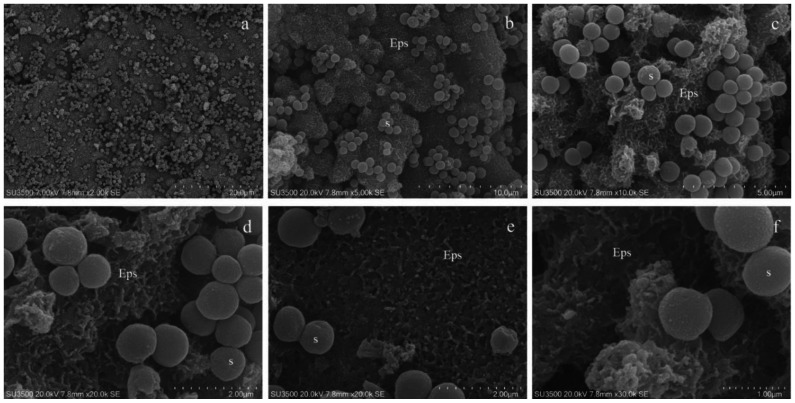
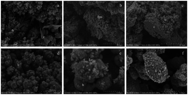
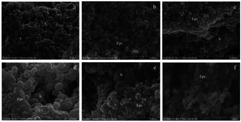
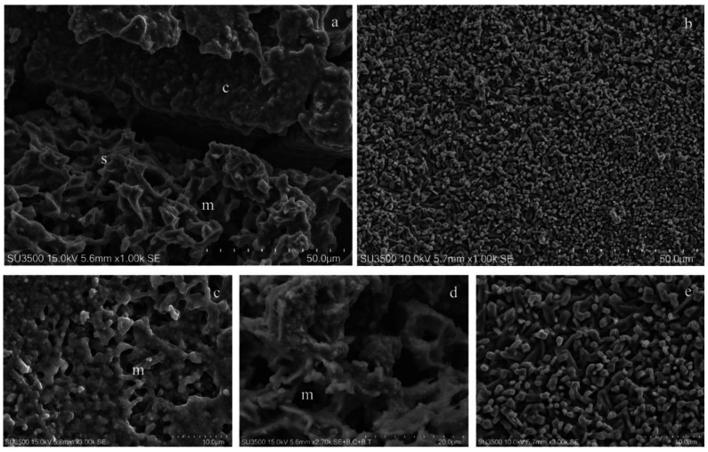
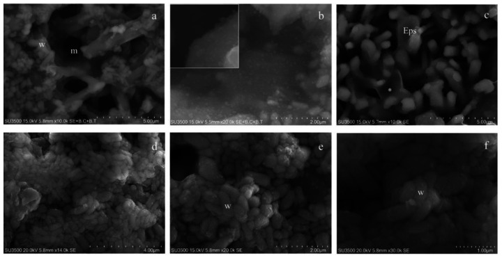
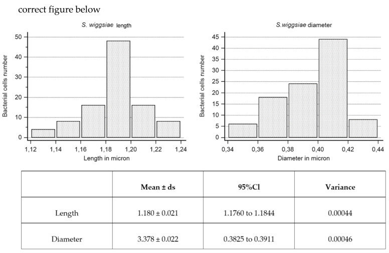
Early childhood caries (ECC) is a severe manifestation of carious pathology with rapid and disruptive progression. The ECC microbiota includes a wide variety of bacterial species, among which is an anaerobic newly named species, Scardovia wiggsiae, a previously unidentified Bifidobacterium. Our aim was to provide the first ultrastructural characterization of S. wiggsiae and its biofilm by scanning electron microscopy (SEM) using a protocol that faithfully preserved the biofilm architecture and allowed an investigation at very high magnifications (order of nanometers) and with the appropriate resolution. To accomplish this task, we analyzed Streptococcus mutans' biofilm by conventional SEM and VP-SEM protocols, in addition, we developed an original procedure, named OsO4-RR-TA-IL, which avoids dehydration, drying and sputter coating. This innovative protocol allowed high-resolution and high-magnification imaging (from 10000× to 35000×) in high-vacuum and high-voltage conditions. After comparing three methods, we chose OsO4-RR-TA-IL to investigate S. wiggsiae. It appeared as a fusiform elongated bacterium, without surface specialization, arranged in clusters and submerged in a rich biofilm matrix, which showed a well-developed micro-canalicular system. Our results provide the basis for the development of innovative strategies to quantify the effects of different treatments, in order to establish the best option to counteract ECC in pediatric patients.

Keywords: Scardovia wiggsiae; Streptococcus mutans scanning electron microscopy; biofilm; early childhood caries; microbiota; pediatric dentistry.


Conflict of interest statement

The authors declare no conflict of interest.

Figures

Figure 1
Figure 1
S. mutans prepared by conventional SEM procedure. (a) SEM, 2000×. At this magnification S. mutans’ biofilm appeared as a compact layer and spherical bacterial cells were scattered on its surface. (b) SEM, 5000×. At increased magnification biofilm surface show spherical bacterial cells arranged in small groups and Eps reveals its rough aspect. Eps: extracellular polymeric substance, S: S. mutans. (c) SEM, 10000×. Eps’s forms a canalicular system of compact trabeculae with a spiny surface. Bacterial cells are adherent to the Eps’s surface. Eps: extracellular polymeric substance, S: S. mutans. (d) SEM, 20000×. Bacterial cells appear irregular, warped and the Eps’s micro-canalicular system is not developed, only superficial holes are visible. Bacterial cells lay down on the Eps’s surface, and they appear naked, without matrix covering. Eps: extracellular polymeric substance, S: S. mutans. (e) SEM, 20000×. Bacterial cells are sometimes fragmented or indented, and the Eps shows a compact aspect due to the collapse of its fine structure. Bacterial cells, uncovered by the matrix, rest on Eps’s surface. Eps: extracellular polymeric substance, S: S. mutans. (f) SEM, 30000×. The highest magnification reveals that the Eps’s layers collapse, causing an obstruction of the Eps’s micro-canalicular system. Bacterial cells appear irregular, on the surface, fine granulation due to sputter coating is visible. Eps: extracellular polymeric substance, S: S. mutans.
Figure 2
Figure 2
S. mutans prepared by conventional VP-SEM procedure. (a) VP-SEM 2000X. Biofilm bacterial towers b forming an intricate micro-canalicular system, and sheet of compact matrix c are shown. (b) VP-SEM 2000×. Spongy biofilm matrix with the Eps showing no sign of collapse or shrinking; it is well preserved in its hydrated state. (c) VP-SEM 5000×. Spongy Eps, s, is developing from a layer of the compact Eps, c. (d) VP-SEM 5000×. Bacterial towers b, superficial granulation represents the Eps secretion. (e) VP-SEM 8000×. On S. mutans’ cells surface, b is clearly visible in the fine graininess of the freshly secreted EPS components. (f) VP-SEM 10000×. Bacterial cell secreting EPS, at this high-magnification image, was vague, and focus is difficult to achieve; these are signs of a low signal to noise ratio, the limit of the VP-SEM technique.
Figure 3
Figure 3
S. mutans prepared by the OsO4-RR-TA-IL procedure. (a) OsO4-RR-TA-IL 3000×. The biofilm topography shows both a compact, c, and spongy, s, appearance. Micro-canalicular system is well developed. (b) OsO4-RR-TA-IL 10000×. At a high magnification, the strength of this technique is revealed, and high-resolution images were obtained without artifacts formation. Eps appears soft, with no signs of collapse or shrinking; micro-canalicular system is intricate. (c) OsO4-RR-TA-IL10000×. Bacterial cells b appeared surrounded by the Eps, spherical and smooth: no shape alterations were found. (d) OsO4-RR-TA-IL10000× Bacterial cells b are completely embedded in the Eps. (e) OsO4-RR-TA-IL15000×. High-voltage, high-magnification and high-resolution image of fully hydrated biofilm, where a single bacterial cell b is partially embedded in the Eps. (f) OsO4-RR-TA-IL 30000×. Very high magnification image confirms the value of this protocol in terms of biofilm three-dimensional structure preservation, until the nanometric level.
Figure 4
Figure 4
Characterization of S. wiggsiae’s biofilm ultrastructural morphology. (a) At a low magnification, 1000×, biofilm shows a compact aspect on the top of the picture, c, spongy on the bottom, s, and a microcanalicular system is evident, m. (b) Biofilm surface shows a fine granular appearance, 1000×. (c) At higher magnifications, a more detailed architecture was distinguishable, compact areas were punctuated with holes, the openings of the microcanalicular system, m. 3000×. (d) Spongy areas reveal a trabecular architecture in whose spaces are a canalicular system, m, and unfold, 2700×. (e) The granular areas were uneven surfaces due to the presence of digitiform extroversions of the biofilm matrix, 3000×.
Figure 5
Figure 5
Characterization of S. wiggsiae bacterial cells. (a) At a very high magnification, 10000×, the biofilm trabecular areas appear populated by clusters of bacteria, w, S. wiggsiae, m, microcanalicular system. (b) Fine granular aggregates with a cloudy appearance are scattered onto the matrix surface 10000X (inset 35000×). (c) Thin matrix sheets, asterisk, connect digitiform extroversions in granular areas creating walls of a canalicular system, 10000×. (d) At a very high magnification, bacteria appear as compact clusters of elongated cells without pili or flagella, 14000×. (e) A thin veil of hydrated matrix covers bacteria, w, giving them a cloudy appearance, 20000×. (f) Very high magnification, 30000×, allows the identification of S. wiggsiae bacterial cells, w, as elongated bacteria without surface extroversions.
Figure 6
Figure 6
S. wiggsiae length and diameter values and their statistical analysis.

Similar articles

Cited by

References

    1. Berg J.H., Slayton R.L. Early Childhood Oral Health. 2nd ed. John Wiley & Sons, Inc.; Hoboken, NJ, USA: 2015.
    1. Douglass J.M., Douglass A.B., Silk H.J. A practical guide to infant oral health. Am. Fam. Physician. 2004;70:2113–2120. - PubMed
    1. Martins-Junior P.A., Vieira-Andrade R.G., Correa-Faria P., Oliveira-Ferreira F., Marques L.S., Ramos-Jorge M.L. Impact of early childhood caries on the oral health-related quality of life of preschool children and their parents. Caries Res. 2013;47:211–218. - PubMed
    1. Meyer F., Enax J. Early childhood caries: Epidemiology, aetiology, and prevention. Int. J. Dent. 2018;2018:1415873. doi: 10.1155/2018/1415873. - DOI - PMC - PubMed
    1. Van Houte J., Gibbs G., Butera C. Oral flora of children with “nursing bottle caries”. J. Dent. Res. 1982;61:382–385. doi: 10.1177/00220345820610020201. - DOI - PubMed

LinkOut - more resources