

Clinical-Grade Peptide-Based Inhibition of CK2 Blocks Viability and Proliferation of T-ALL Cells and Counteracts IL-7 Stimulation and Stromal Support
- PMID: 32471246
- PMCID: PMC7352628
- DOI: 10.3390/cancers12061377
Clinical-Grade Peptide-Based Inhibition of CK2 Blocks Viability and Proliferation of T-ALL Cells and Counteracts IL-7 Stimulation and Stromal Support
Abstract
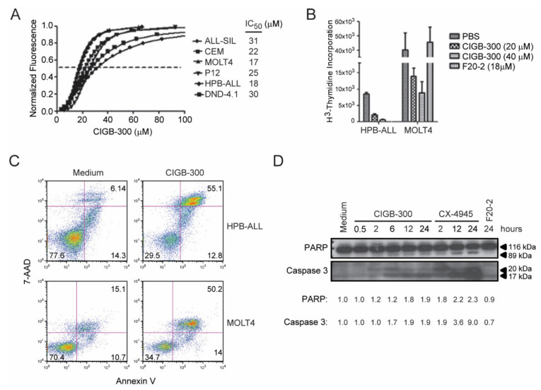
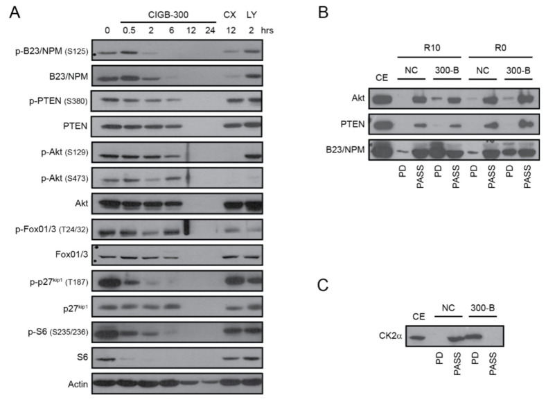
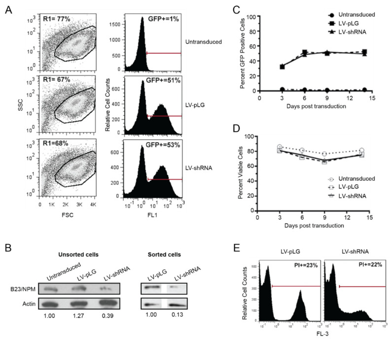
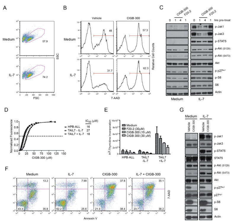
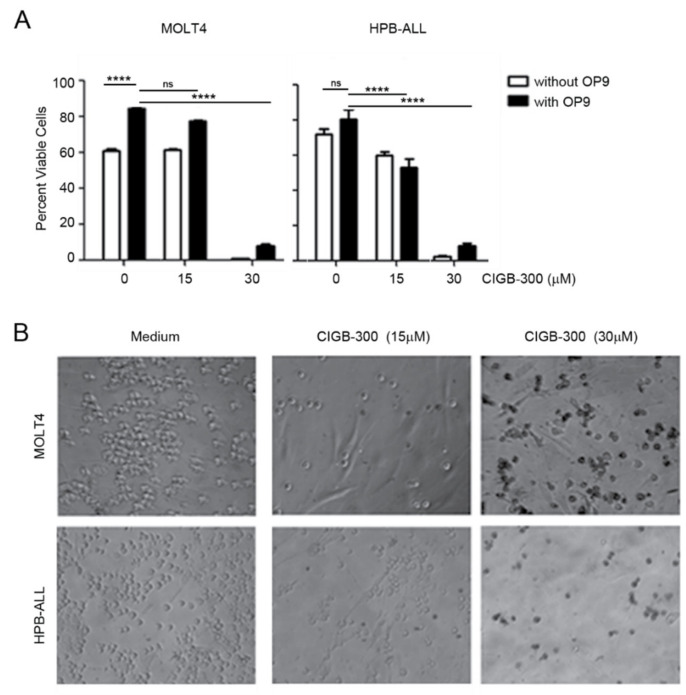
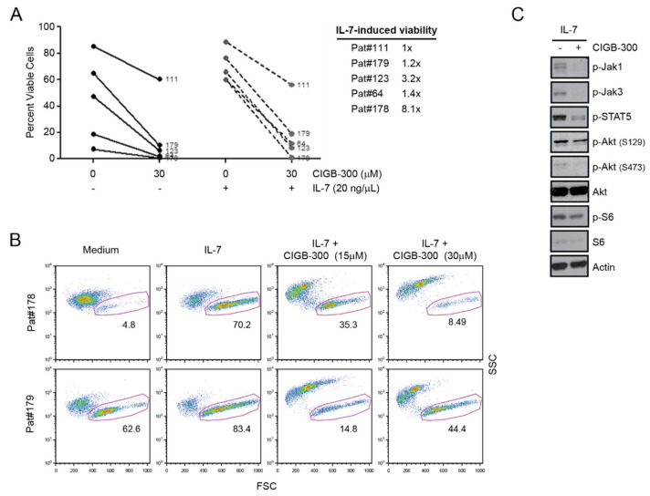
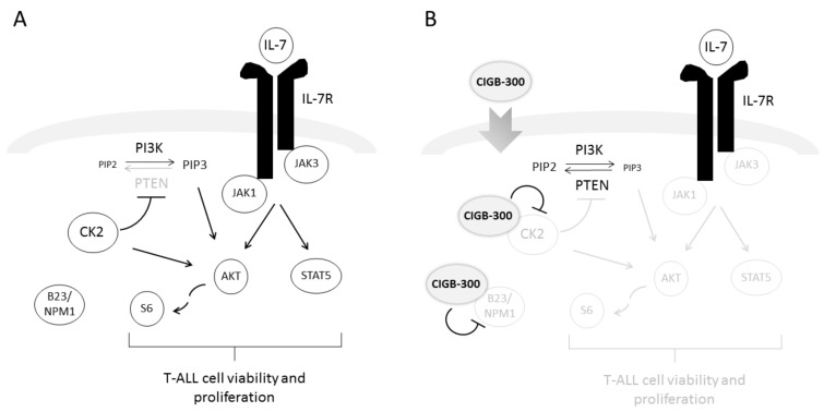
Despite remarkable advances in the treatment of T-cell acute lymphoblastic leukemia (T-ALL), relapsed cases are still a major challenge. Moreover, even successful cases often face long-term treatment-associated toxicities. Targeted therapeutics may overcome these limitations. We have previously demonstrated that casein kinase 2 (CK2)-mediated phosphatase and tensin homologue (PTEN) posttranslational inactivation, and consequent phosphatidylinositol 3-kinase (PI3K)/Akt signaling hyperactivation, leads to increased T-ALL cell survival and proliferation. We also revealed the existence of a crosstalk between CK2 activity and the signaling mediated by interleukin 7 (IL-7), a critical leukemia-supportive cytokine. Here, we evaluated the impact of CIGB-300, a the clinical-grade peptide-based CK2 inhibitor CIGB-300 on T-ALL biology. We demonstrate that CIGB-300 decreases the viability and proliferation of T-ALL cell lines and diagnostic patient samples. Moreover, CIGB-300 overcomes IL-7-mediated T-ALL cell growth and viability, while preventing the positive effects of OP9-delta-like 1 (DL1) stromal support on leukemia cells. Signaling and pull-down experiments indicate that the CK2 substrate nucleophosmin 1 (B23/NPM1) and CK2 itself are the molecular targets for CIGB-300 in T-ALL cells. However, B23/NPM1 silencing only partially recapitulates the anti-leukemia effects of the peptide, suggesting that CIGB-300-mediated direct binding to CK2, and consequent CK2 inactivation, is the mechanism by which CIGB-300 downregulates PTEN S380 phosphorylation and inhibits PI3K/Akt signaling pathway. In the context of IL-7 stimulation, CIGB-300 blocks janus kinase / signal transducer and activator of transcription (JAK/STAT) signaling pathway in T-ALL cells. Altogether, our results strengthen the case for anti-CK2 therapeutic intervention in T-ALL, demonstrating that CIGB-300 (given its ability to circumvent the effects of pro-leukemic microenvironmental cues) may be a valid tool for clinical intervention in this aggressive malignancy.
Keywords: CIGB-300; Casein kinase 2 (CK2); IL-7 receptor (IL-7R); IL-7-mediated signaling; Signaling therapies.; Stromal support; T-cell acute lymphoblastic leukemia (T-ALL).
Conflict of interest statement
The authors declare no conflict of interest.
Figures
Similar articles
-
Targeting of Protein Kinase CK2 in Acute Myeloid Leukemia Cells Using the Clinical-Grade Synthetic-Peptide CIGB-300.Biomedicines. 2021 Jul 1;9(7):766. doi: 10.3390/biomedicines9070766. Biomedicines. 2021. PMID: 34356831 Free PMC article.
-
Anticancer peptide CIGB-300 binds to nucleophosmin/B23, impairs its CK2-mediated phosphorylation, and leads to apoptosis through its nucleolar disassembly activity.Mol Cancer Ther. 2009 May;8(5):1189-96. doi: 10.1158/1535-7163.MCT-08-1056. Epub 2009 May 5. Mol Cancer Ther. 2009. PMID: 19417160
-
New insights into Notch1 regulation of the PI3K-AKT-mTOR1 signaling axis: targeted therapy of γ-secretase inhibitor resistant T-cell acute lymphoblastic leukemia.Cell Signal. 2014 Jan;26(1):149-61. doi: 10.1016/j.cellsig.2013.09.021. Epub 2013 Oct 16. Cell Signal. 2014. PMID: 24140475 Review.
-
CIGB-300 internalizes and impairs viability of NSCLC cells lacking actionable targets by inhibiting casein kinase-2 signaling.Sci Rep. 2024 Oct 29;14(1):26038. doi: 10.1038/s41598-024-75990-1. Sci Rep. 2024. PMID: 39472715 Free PMC article.
-
Casein Kinase II (CK2), Glycogen Synthase Kinase-3 (GSK-3) and Ikaros mediated regulation of leukemia.Adv Biol Regul. 2017 Aug;65:16-25. doi: 10.1016/j.jbior.2017.06.001. Epub 2017 Jun 13. Adv Biol Regul. 2017. PMID: 28623166 Free PMC article. Review.
Cited by
-
CIGB-300-Regulated Proteome Reveals Common and Tailored Response Patterns of AML Cells to CK2 Inhibition.Front Mol Biosci. 2022 Mar 11;9:834814. doi: 10.3389/fmolb.2022.834814. eCollection 2022. Front Mol Biosci. 2022. PMID: 35359604 Free PMC article.
-
CIGB-300 Peptide Targets the CK2 Phospho-Acceptor Domain on Human Papillomavirus E7 and Disrupts the Retinoblastoma (RB) Complex in Cervical Cancer Cells.Viruses. 2022 Jul 30;14(8):1681. doi: 10.3390/v14081681. Viruses. 2022. PMID: 36016303 Free PMC article.
-
Targeting of Protein Kinase CK2 Elicits Antiviral Activity on Bovine Coronavirus Infection.Viruses. 2022 Mar 7;14(3):552. doi: 10.3390/v14030552. Viruses. 2022. PMID: 35336959 Free PMC article.
-
CIGB-300 Anticancer Peptide Differentially Interacts with CK2 Subunits and Regulates Specific Signaling Mediators in a Highly Sensitive Large Cell Lung Carcinoma Cell Model.Biomedicines. 2022 Dec 25;11(1):43. doi: 10.3390/biomedicines11010043. Biomedicines. 2022. PMID: 36672551 Free PMC article.
-
Targeting of Protein Kinase CK2 in Acute Myeloid Leukemia Cells Using the Clinical-Grade Synthetic-Peptide CIGB-300.Biomedicines. 2021 Jul 1;9(7):766. doi: 10.3390/biomedicines9070766. Biomedicines. 2021. PMID: 34356831 Free PMC article.
References
-
- Silva A., Yunes J.A., Cardoso B.A., Martins L.R., Jotta P.Y., Abecasis M., Nowill A.E., Leslie N.R., Cardoso A.A., Barata J.T. PTEN posttranslational inactivation and hyperactivation of the PI3K/Akt pathway sustain primary T cell leukemia viability. J. Clin. Investig. 2008;118:3762–3774. doi: 10.1172/JCI34616. - DOI - PMC - PubMed
-
- Oliveira M.L., Akkapeddi P., Alcobia I., Almeida A.R., Cardoso B.A., Fragoso R., Serafim T.L., Barata J.T. From the outside, from within: Biological and therapeutic relevance of signal transduction in T-cell acute lymphoblastic leukemia. Cell. Signal. 2017;38:10–25. doi: 10.1016/j.cellsig.2017.06.011. - DOI - PubMed
Grants and funding
LinkOut - more resources
Full Text Sources
Other Literature Sources
Molecular Biology Databases
Research Materials
