. 2020 May 27;21(11):3811.
doi: 10.3390/ijms21113811.

The FT-IR and Raman Spectroscopies as Tools for Biofilm Characterization Created by Cariogenic Streptococci

Affiliations

The FT-IR and Raman Spectroscopies as Tools for Biofilm Characterization Created by Cariogenic Streptococci

Barbara Gieroba et al. Int J Mol Sci. .

Abstract

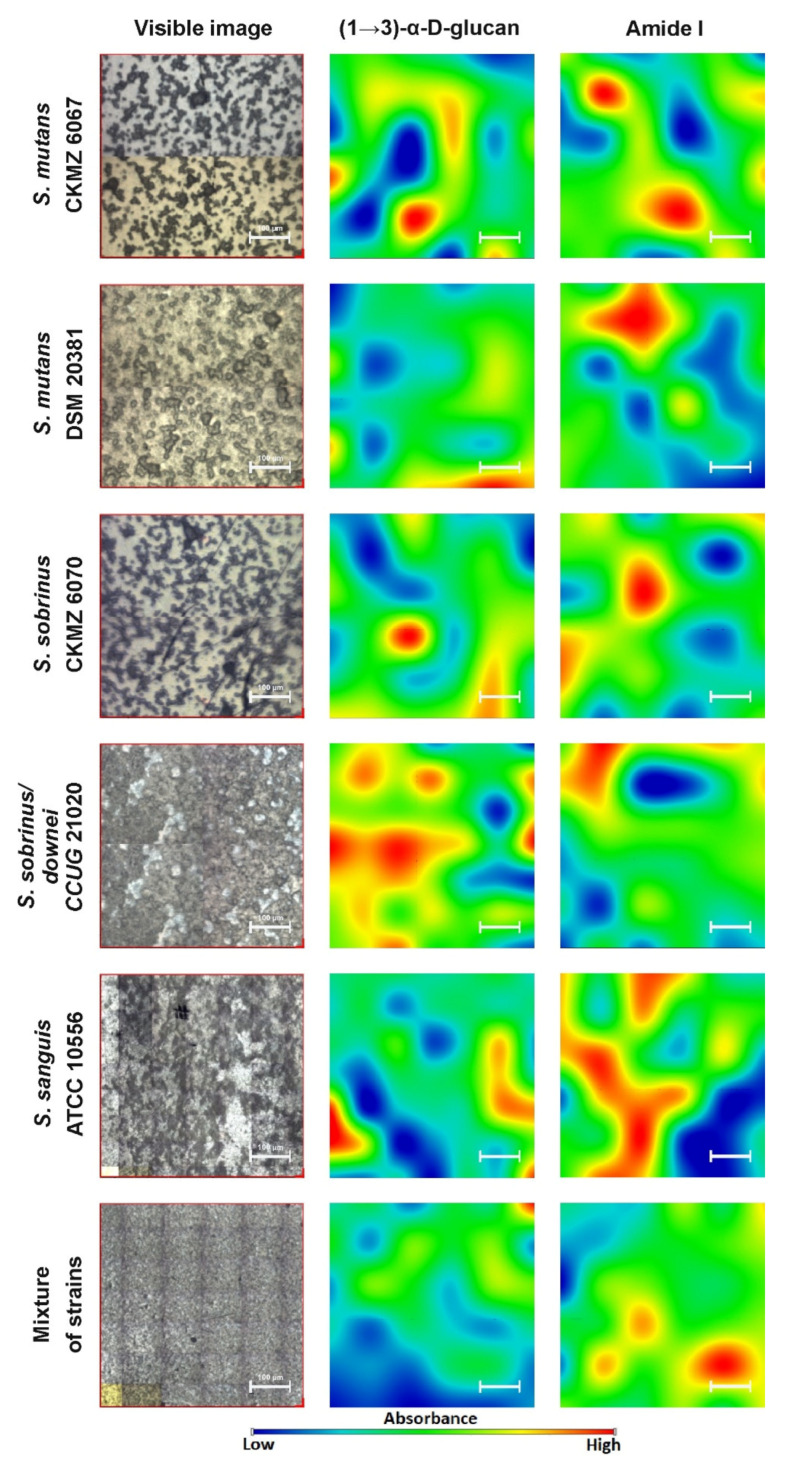
Fourier transform infrared (FT-IR) and Raman spectroscopy and mapping were applied to the analysis of biofilms produced by bacteria of the genus Streptococcus. Bacterial biofilm, also called dental plaque, is the main cause of periodontal disease and tooth decay. It consists of a complex microbial community embedded in an extracellular matrix composed of highly hydrated extracellular polymeric substances and is a combination of salivary and bacterial proteins, lipids, polysaccharides, nucleic acids, and inorganic ions. This study confirms the value of Raman and FT-IR spectroscopies in biology, medicine, and pharmacy as effective tools for bacterial product characterization.

Keywords: FT-IR microspectroscopy; Raman spectroscopy; bacteria; bacterial polysaccharides; biofilms; dental caries; mutans streptococci.


Conflict of interest statement

The authors declare no conflict of interest. The funders had no role in the design of the study, in the collection, analyses, or interpretation of data, in the writing of the manuscript, or in the decision to publish the results.

Figures

Figure 1
Figure 1
Representative relative intensity of Fourier transform infrared (FT-IR) spectra of bacterial biofilms.
Figure 2
Figure 2
The FT-IR spectra of bacterial biofilms normalized to the Amide I band.
Figure 3
Figure 3
The relative intensity of FT-IR spectra of the fingerprints regions of: (A)—lipid region (3000–2800 cm−1), (B)—Amides I and II region (1700–1470 cm−1), and (C)—carbohydrate region (1200–700 cm−1).
Figure 4
Figure 4
The second-order derivatives of the FT-IR spectra of biofilms; (A)—lipid region; (B)—protein region; (C)—carbohydrate region.
Figure 5
Figure 5
The FT-IR chemical maps of compound distributions in bacterial biofilms. The white bar corresponds to 100 µm.
Figure 6
Figure 6
Representative relative intensity of the Raman spectra of bacterial biofilms.
Figure 7
Figure 7
The Raman spectra normalized to the 1300–1400 cm−1 band (assigned to deformation vibration of the CH2 group in lipids and proteins) of bacterial biofilms.
Figure 8
Figure 8
The relative intensity of the Raman spectra of the fingerprint region spectra of: (A)—lipid and Amide III region (1500–1175 cm−1), (B)—Amides I and II region (1750–1500 cm−1), and (C)—carbohydrate region (1200–800 and 610–475 cm−1).
Figure 9
Figure 9
The second-order derivatives of the Raman spectra of biofilms; (A)—lipid region; (B)—protein region; (C)—carbohydrate region.
Figure 10
Figure 10
The Raman chemical maps of compound distributions in bacterial biofilms. The red cross and frame in the optical images indicate the mapping area. The white bar in the chemical maps corresponds to 1 µm.

References

    1. Marsh P.D. Dental diseases—Are these examples of ecological catastrophes? Int. J. Dent. Hyg. 2006;4(Suppl. 1):3–10. doi: 10.1111/j.1601-5037.2006.00195.x. - DOI - PubMed
    1. Pleszczyńska M., Wiater A., Janczarek M., Szczodrak J. (1→3)-α-d-Glucan hydrolases in dental biofilm prevention and control: A review. Int. J. Biol. Macromol. 2015;79:761–778. doi: 10.1016/j.ijbiomac.2015.05.052. - DOI - PubMed
    1. Loesche W.J. Role of Streptococcus mutans in human dental decay. Microbiol. Rev. 1986;50:353–380. doi: 10.1128/MMBR.50.4.353-380.1986. - DOI - PMC - PubMed
    1. Aoki H., Shiroza T., Hayakawa M., Sato S., Kuramitsu H.K. Cloning of a streptococcus mutans glucosyltransferase gene coding for insoluble glucan synthesis. Infect. Immun. 1986;53:587–594. doi: 10.1128/IAI.53.3.587-594.1986. - DOI - PMC - PubMed
    1. Hanada N., Kuramitsu H.K. Isolation and characterization of the Streptococcus mutans gtfD gene, coding for primer-dependent soluble glucan synthesis. Infect. Immun. 1989;57:2079–2085. doi: 10.1128/IAI.57.7.2079-2085.1989. - DOI - PMC - PubMed

MeSH terms

Substances