Review
. 2020 May 27;9(6):1341.
doi: 10.3390/cells9061341.

Endothelial TRPV1 as an Emerging Molecular Target to Promote Therapeutic Angiogenesis

Affiliations
Review

Endothelial TRPV1 as an Emerging Molecular Target to Promote Therapeutic Angiogenesis

Sharon Negri et al. Cells. .

Abstract

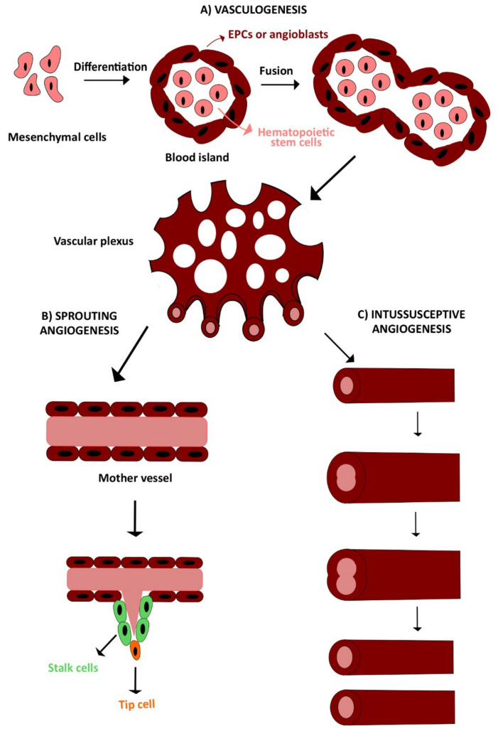
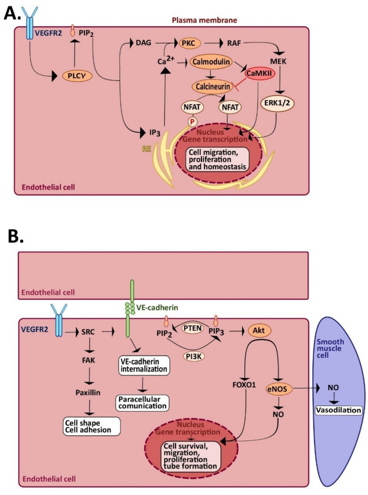
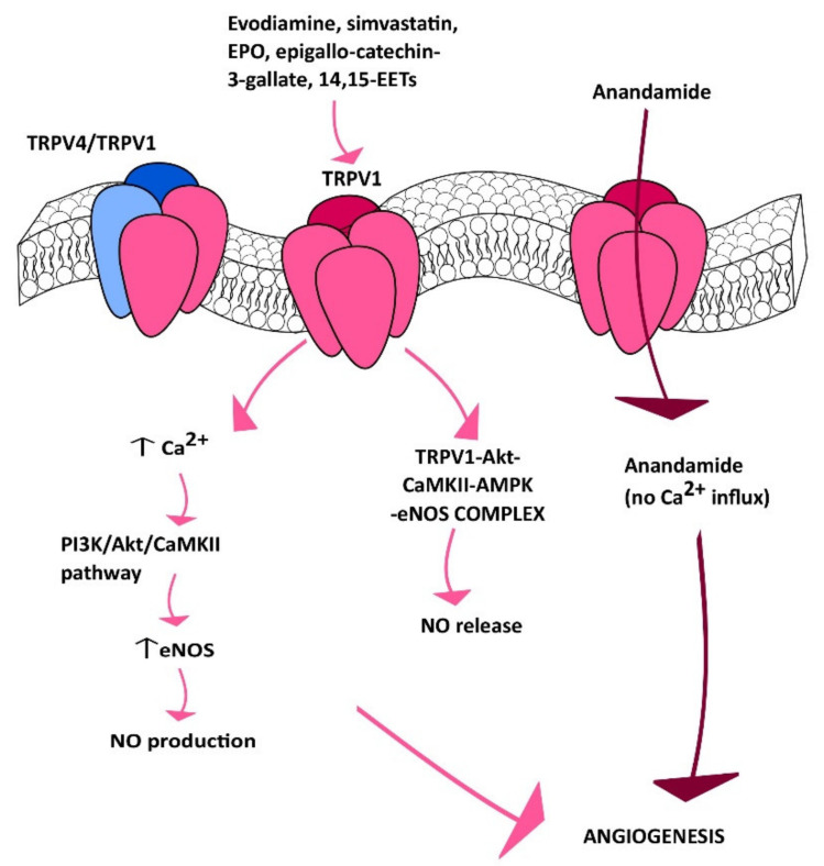
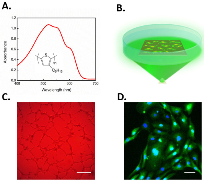
Therapeutic angiogenesis represents an emerging strategy to treat ischemic diseases by stimulating blood vessel growth to rescue local blood perfusion. Therefore, injured microvasculature may be repaired by stimulating resident endothelial cells or circulating endothelial colony forming cells (ECFCs) or by autologous cell-based therapy. Endothelial Ca2+ signals represent a crucial player in angiogenesis and vasculogenesis; indeed, several angiogenic stimuli induce neovessel formation through an increase in intracellular Ca2+ concentration. Several members of the Transient Receptor Potential (TRP) channel superfamily are expressed and mediate Ca2+-dependent functions in vascular endothelial cells and in ECFCs, the only known truly endothelial precursor. TRP Vanilloid 1 (TRPV1), a polymodal cation channel, is emerging as an important player in endothelial cell migration, proliferation, and tubulogenesis, through the integration of several chemical stimuli. Herein, we first summarize TRPV1 structure and gating mechanisms. Next, we illustrate the physiological roles of TRPV1 in vascular endothelium, focusing our attention on how endothelial TRPV1 promotes angiogenesis. In particular, we describe a recent strategy to stimulate TRPV1-mediated pro-angiogenic activity in ECFCs, in the presence of a photosensitive conjugated polymer. Taken together, these observations suggest that TRPV1 represents a useful target in the treatment of ischemic diseases.

Keywords: Ca2+ signaling; TRPV1; endothelial colony forming cells; erythropoietin; evodiamine; organic semiconductors; photostimulation; simvastatin; therapeutic angiogenesis; vascular endothelial cells.


Conflict of interest statement

The authors declare no conflict of interest.

Figures

Figure 1
Figure 1
The two main processes involved in vascular development: vasculogenesis and angiogenesis. (A) Schematic representation of vasculogenesis, which consists of de novo vessel formation from aggregated endothelial precursors (EPCs or angioblasts) assembled within the blood islands. Thereafter, multiple blood islands fuse together into the early vascular plexus, which in turn generates primitive blood vessels. (B,C) Angiogenesis consists of neovessel formation from preexisting blood vessels in response to pro-angiogenic signals. Angiogenesis may occur through two different mechanisms: sprouting angiogenesis and intussusceptive angiogenesis. See text for detailed explanation.
Figure 2
Figure 2
Role of endothelial Ca2+ signaling angiogenesis and vasculogenesis. (A) VEGF binds to VEGFR2 and induces receptor dimerization and transphosphorylation. Activation of VEGFR2, in turn, leads to InsP3-dependent Ca2+ release, followed by SOCE activation (not shown). The ensuing increase in [Ca2+]i leads to the activation of Ca2+-dependent decoders, such as calmodulin, which in turn activates calcineurin and Ca2+-Calmodulin-dependent kinase II (CaMKII). Calcineurin dephosphorylates NFAT, thereby promoting its translocation into the nucleus, where it activates genes responsible for cell proliferation and migration. CaMKII, in turn, inhibits calcineurin activity, but stimulates angiogenesis by phosphorylating multiple targets (i.e., Akt and Src, not shown). In addition, the increase in [Ca2+]i promotes cytosolic protein kinase C (PKC) relocation toward the plasma membrane. Herein, PKC is activated by DAG to stimulate the extracellular signal-regulated kinases 1/2 (ERK1/2) phosphorylation cascade. (B) Schematic representation of the Src and PI3K-Akt signaling pathways activated by VEGF. Once activated, VEGFR2 activates Src and subsequent downstream pathways involved in cell shape, adhesion, permeability, and survival. Moreover, VEGFR2 indirectly activates PI3K, either by Src or by VE-cadherin. PI3K generates phosphatidylinositol-3,4,5-trisphosphate (PIP3), which activates Akt, followed by eNOS activation and NO release. Finally, Akt activates FOXO1, which translocates into the nucleus, where, together with NO, induces transcription of genes involved in cell survival, migration, proliferation, and tube formation.
Figure 3
Figure 3
TRPV1 channel in angiogenesis. TRPV1 stimulates angiogenesis in response to evodiamine, simvastatin, EPO, epigallo-catechin-3-gallate, and 14,15-EETS in a Ca2+-dependent manner. Conversely, extracellular anandamide may enter through TRPV1, thereby stimulating angiogenesis in a Ca2+-independent manner.
Figure 4
Figure 4
Proposed molecular mechanism of eNOS stimulation after TRPV1 activation. Activation of TRPV1 increases Ca2+ influx, which in turn activates PI3K/Akt/CaMKII signaling, leading to increased TRPV1 and eNOS phosphorylation. In addition, TRPV1 may serve as a scaffold for the formation of a complex comprising Akt, AMPK, CaMKII, and eNOS. Protein interactions seem to be important in eNOS activation and NO release.
Figure 5
Figure 5
(A) The rr-P3HT chemical structure and optical absorption of the thin film. (B) Experimental setup and optical excitation protocol performed for evaluation of polymer-mediated cell photoexcitation effects on cell fate. (C) Representative image of in vitro network formation in ECFCs seeded on P3HT and subjected to optical excitation; scale bar: 250 µM. (D) Representative image of immunofluorescence staining, showing enhanced light-induced NF-κB nuclear translocation. Cell nuclei are detected by DAPI (blue) while cytoplasmic p65 NF-κB subunit with a secondary chicken anti-rabbit Alexa(488)-conjugated antibody (green). Scale bar: 50 µM. Figures adapted from [44,207].

Similar articles

Cited by

References

    1. McCarron J.G., Lee M.D., Wilson C. The Endothelium Solves problems that endothelial cells do not know exist. Trends Pharmacol. Sci. 2017;38:322–338. doi: 10.1016/j.tips.2017.01.008. - DOI - PMC - PubMed
    1. McCarron J.G., Wilson C., Heathcote H.R., Zhang X., Buckley C., Lee M.D. Heterogeneity and emergent behaviour in the vascular endothelium. Curr. Opin. Pharmacol. 2019;45:23–32. doi: 10.1016/j.coph.2019.03.008. - DOI - PMC - PubMed
    1. Carmeliet P., Jain R.K. Molecular mechanisms and clinical applications of angiogenesis. Nature. 2011;473:298–307. doi: 10.1038/nature10144. - DOI - PMC - PubMed
    1. Banno K., Yoder M.C. Tissue regeneration using endothelial colony-forming cells: Promising cells for vascular repair. Pediatr. Res. 2018;83:283–290. doi: 10.1038/pr.2017.231. - DOI - PubMed
    1. Paschalaki K.E., Randi A.M. Recent Advances in endothelial colony forming cells toward their use in clinical translation. Front. Med. 2018;5:295. doi: 10.3389/fmed.2018.00295. - DOI - PMC - PubMed

Publication types

LinkOut - more resources