. 2020 May 29;21(1):132.
doi: 10.1186/s12931-020-01384-2.

TRIM72 promotes alveolar epithelial cell membrane repair and ameliorates lung fibrosis

Affiliations

TRIM72 promotes alveolar epithelial cell membrane repair and ameliorates lung fibrosis

Xiaofei Cong et al. Respir Res. .

Abstract

Background: Chronic tissue injury was shown to induce progressive scarring in fibrotic diseases such as idiopathic pulmonary fibrosis (IPF), while an array of repair/regeneration and stress responses come to equilibrium to determine the outcome of injury at the organ level. In the lung, type I alveolar epithelial (ATI) cells constitute the epithelial barrier, while type II alveolar epithelial (ATII) cells play a pivotal role in regenerating the injured distal lungs. It had been demonstrated that eukaryotic cells possess repair machinery that can quickly patch the damaged plasma membrane after injury, and our previous studies discovered the membrane-mending role of Tripartite motif containing 72 (TRIM72) that expresses in a limited number of tissues including the lung. Nevertheless, the role of alveolar epithelial cell (AEC) repair in the pathogenesis of IPF has not been examined yet.

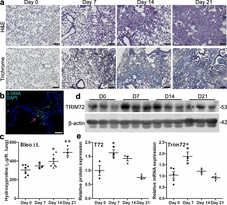
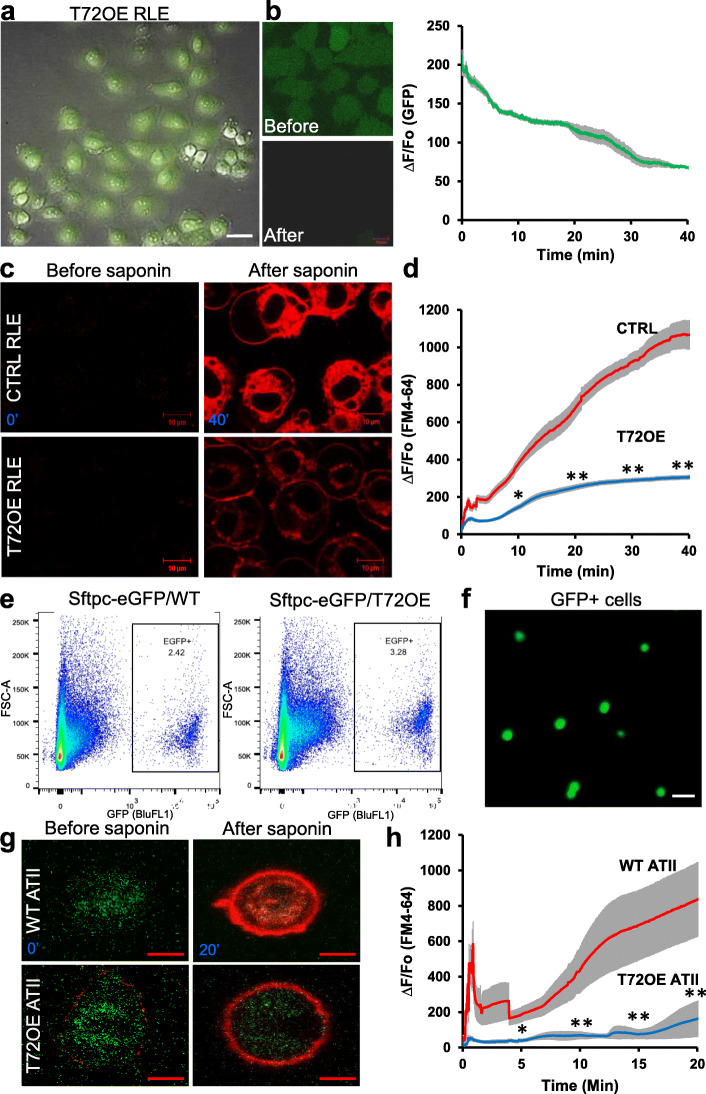
Method: In this study, we tested the specific roles of TRIM72 in the repair of ATII cells and the development of lung fibrosis. The role of membrane repair was accessed by saponin assay on isolated primary ATII cells and rat ATII cell line. The anti-fibrotic potential of TRIM72 was tested with bleomycin-treated transgenic mice.

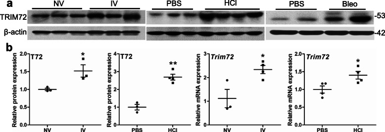
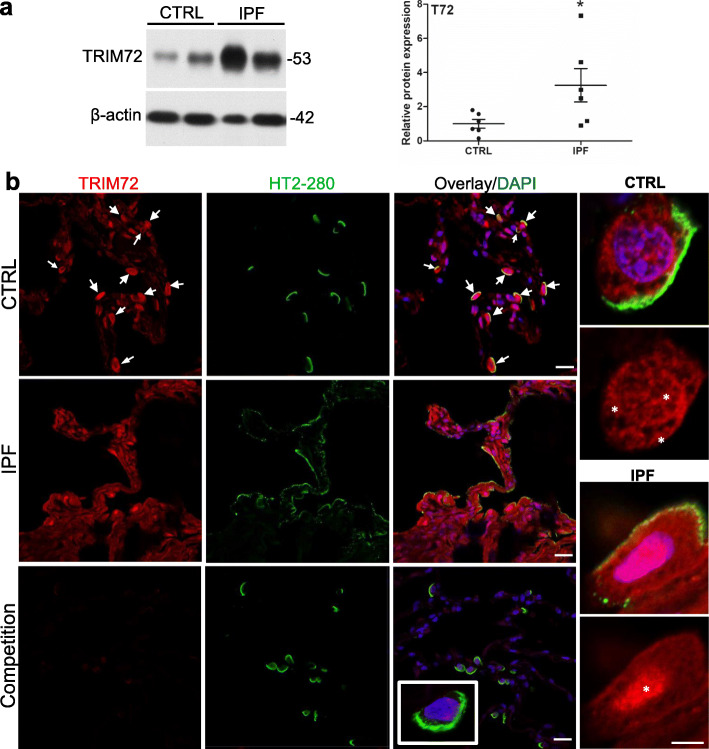
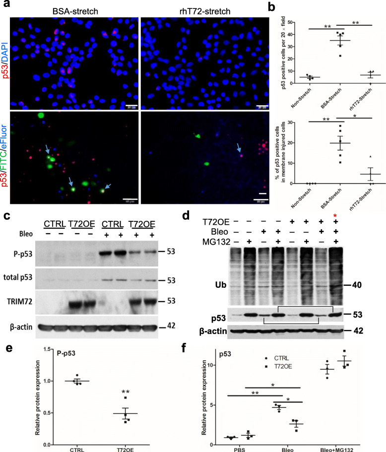
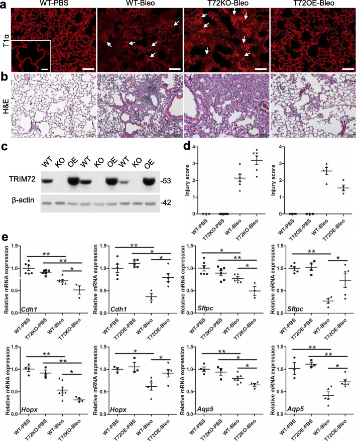
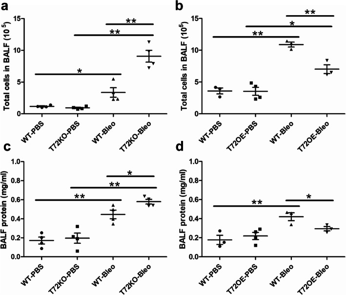
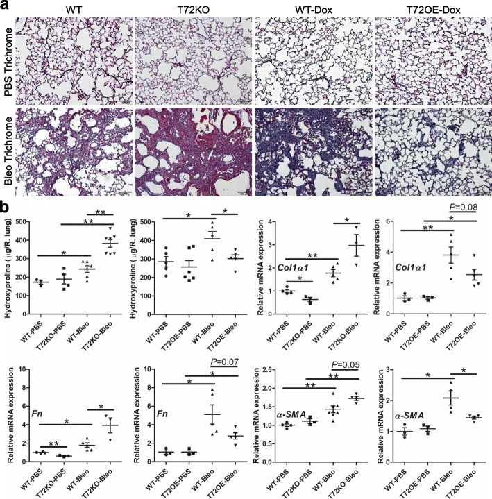
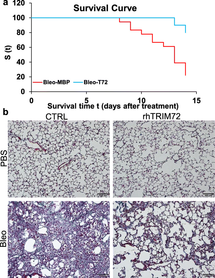
Results: We showed that TRIM72 was upregulated following various injuries and in human IPF lungs. However, TRIM72 expression in ATII cells of the IPF lungs had aberrant subcellular localization. In vitro studies showed that TRIM72 repairs membrane injury of immortalized and primary ATIIs, leading to inhibition of stress-induced p53 activation and reduction in cell apoptosis. In vivo studies demonstrated that TRIM72 protects the integrity of the alveolar epithelial layer and reduces lung fibrosis.

Conclusion: Our results suggest that TRIM72 protects injured lungs and ameliorates fibrosis through promoting post-injury repair of AECs.

Keywords: Alveolar epithelial cells; Apoptosis; Idiopathic pulmonary fibrosis; Membrane repair; Tripartite motif family protein 72.


Conflict of interest statement

The authors declare that they have no competing interests.

Figures

Fig. 1
Fig. 1
TRIM72 expression in the lung is induced by various injurious stimuli. a Western blot of TRIM72 protein in wildtype (WT) lungs received normal tidal volume (NV) or injurious ventilation (IV) for 3 h, intra-tracheal (i.t.) injection of PBS, 0.1 N hydrochloride acid (HCl) or 2 U/kg bleomycin (bleo). Tissues were harvested from mice 24 h after HCl i.t. or 14 d after bleo i.t.. The molecular weight (kDa) of proteins was labeled on all Western images; b quantification of TRIM72 protein or Trim72 mRNA in IV vs. NV or HCl vs PBS lungs. Band intensity is normalized to β-actin and shown in mean ± SEM, n = 3 for NV and IV, n = 3 for PBS, n = 4 for HCl, *P < 0.05, **P < 0.01 compared to NV or PBS based on two-tailed student t-test
Fig. 2
Fig. 2
TRIM72 protein expression and distribution in the IPF lung. a Western blot and quantification of TRIM72 protein in histologically normal para-tumor (control, CTRL) human lung specimens and pathologically confirmed idiopathic pulmonary fibrosis (IPF) lung specimens. n = 6 for CTRL or IPF groups; b immunostaining of TRIM72 and HT2–280 on CTRL and IPF human lung sections. HT2–280 is a membrane-bound marker for human type II alveolar epithelial cells (ATII). Competitive immunostaining using 10 μg/ml recombinant human TRIM72 protein (rhT72) was included as a control for staining specificity of the anti-human TRIM72 antibody. White arrows = TRIM72 positive ATII cells; asterisks = cellular location of TRIM72. Scale bar = 20 μm for full images, = 5 μm for high magnification images
Fig. 3
Fig. 3
Tapering of bleomycin (bleo)-induced TRIM72 upregulation correlates with an increase in lung collagen content. a H&E and Masson’s trichrome staining on lung sections from 5 to 6-month-old B6 WT mice received i.t. of 75 μl 1.5 U/kg bleo. Tissues were harvested at days 0, 7, 14, and 21 after i.t. respectively. Scale bar = 100 μm; b α-smooth muscle actin immunostaining (α-SMA, green) on bleo-treated lung, counter-stained with DAPI (blue). Red arrow = fibrotic area. Scale bar = 50 μm; c hydroxyproline content per right lung after bleo i.t.; d Western blot of TRIM72 in bleo-treated WT lungs at the above time points; e quantification of TRIM72 protein and Trim72 mRNA in mouse lung samples. n = 4 for D0 and D7, n = 6 for D14 and D21, *P < 0.05 or **P < 0.01 compared to day 0 based on one-way ANOVA with post hoc analysis
Fig. 4
Fig. 4
TRIM72 promotes membrane repair of ATII-like rat lung epithelial cells (RLE). a Lentivirus-mediated TRIM72 expression (T72OE) in RLE cells. Expression of GFP marker on the L309 vector indicates infected cells; b 0.005% saponin injury releases intracellular GFP from RLE cells; c representative images of FM4–64 dye entry in CTRL and T72OE RLE cells before (0′) and after saponin treatment (40′). Scale bar = 10 μm; d quantification of FM4–64 dye entry normalized to baseline fluorescence (∆F/F0). n = 36 cells for (CTRL RLE and n = 39 for T72OE RLE cells. *P < 0.05 or **P < 0.01 compared to WT at 10′, 20′, 30′ and 40′ based on two-sided student t-tests; e Flow sorting of GFP+ primary ATII cells from lungs of the sftpc-eGFP/WT and sftpc-eGFP/TRIM72 overexpressor (T72OE) mice; f representative image of freshly sorted GFP-positive primary ATII cells; scale bar = 50 μm; g representative images of FM4–64 dye entry in primary WT and T72OE ATII cells before (0′) and after saponin treatment (20′). Scale bar = 10 μm; h quantification of FM4–64 dye entry normalized to baseline fluorescence (∆F/F0). n = 8 cells for WT ATII and n = 4 for T72OE ATII cells. *P < 0.05, or **P < 0.01 compared to WT at 5′, 10′, 15′ and 20′ based on two-tailed student t-tests
Fig. 5
Fig. 5
TRIM72 salvages stress-induced p53 activation in vitro. a Upper panels: immunostaining of total p53 (red) on stretch-injured RLE cells treated with bovine serum albumin (BSA) or rhT72; cells are counter-stained with DAPI (blue); lower panels: representative images of repaired cells labeled with FITC-dextran (green), non-repaired cells labeled with fixable cell vitality dye eFluor450 (blue) and post-fixation immunostaining of p53 (red) in these cells; blue arrows = p53+ FITC+ or p53 + eFluor+ cells; scale bar = 20 μm; b the number of p53 positive cells per 20 × field and quantification of p53+ cells among non-stretched and membrane injured cells (FITC+ plus eFluor+); c Western blot of Ser15 phosphorylated p53 (P-p53), total p53, and TRIM72 in CTRL or T72OE RLE cells with or without treatment of 50 μg/ml bleo; b Western blot detection of ubiquitin and total p53 in T72OE or CTRL, in the presence and absence of bleo and with or without MG132 to inhibit proteasome degradation of ubiquitinated substrates. Stars: Bleo+MG132-treated CTRL and T72OE RLE cells; brackets: bleo-induced total p53 with or without MG132 treatment; c and d quantification of P-p53 and p53 (in the absence and presence of MG132) from bleo-treated RLE cells. Relative protein expression of P-p53 or p53 was normalized to β-actin. n = 4, *P < 0.05, or **P < 0.01 based on two-tailed student t-test (P-p53) or one-way ANOVA with post hoc analysis (p53)
Fig. 6
Fig. 6
TRIM72 inhibits bleomycin (bleo)-induced ATII cell apoptosis. a Representative TUNEL, SFTPC, and cleaved caspase-3 (Casp-3) immunostaining on doxycycline (Dox)-injected bleo-treated WT and TRIM72 overexpressor (T72OE) lungs; scale bar = 100 μm; b quantification of the number of apoptotic cells (TUNEL+); c percentage of apoptotic ATII cells (SFTPC positive) among TUNEL positive cells, and apoptotic ATII cells among total ATII cells in PBS- or bleo-treated lungs; data = mean ± SEM, n = 3 for WT and T72OE PBS and n = 6 for WT and T72OE bleo groups, *P < 0.05 or **P < 0.01 compared to WT based on two-tailed student t-tests, or one-way ANOVA with post hoc analysis for comparison among different groups
Fig. 7
Fig. 7
TRIM72 maintains alveolar epithelial integrity in stressed lungs. a Immunostaining of T1α to indicate alveolar epithelial integrity in PBS- and bleo-treated B6 WT, T72KO, and T72OE (Dox injected) lungs. Scale bar = 100 μm; b H&E staining of bleo-treated WT, T72KO, and T72OE lungs. Scale bar = 100 μm. The lungs from 2 to 3-month-old B6 WT and 5–6-month-old Dox-injected 129/B6 WT mice showed no difference in immunostaining of T1α or H&E staining; c. validation of experimental models. a Western blot shows good efficiency of Dox-induced TRIM72OE transgene induction and lack of TRIM72 expression in the TRIM72 knockout (KO) lungs; d injury scores based on T1α-staining indicated epithelial disruption. Mann Whitney U test was used since the injury scores are non-parametric data. The results indicated statistically significant differences; e relative mRNA expression levels of Cdh1 (E-cadherin), sftpc, Hopx, and Aqp5 in bleo-treated lungs, n = 4 for PBS groups and n = 6 for bleo groups; *P < 0.05 or **P < 0.01 compared to WT groups based on two-sided student t-tests
Fig. 8
Fig. 8
BAL fluid (BALF) cell profiles and BALF protein of bleo-injured lungs. a total BALF cells in WT vs. T72KO and b WT vs. T72OE lungs at day 3 after bleo i.t.; c BALF protein in WT vs. T72KO and d WT vs. T72OE lungs at day 3 after bleo i.t.. n = 4 for PBS and bleo groups, *P < 0.05 or **P < 0.01 based on one-way ANOVA with post hoc analysis
Fig. 9
Fig. 9
TRIM72 protects bleomycin (bleo)-induced lung injury and fibrosis. a Masson’s trichrome staining in bleo-treated B6 WT, T72KO, Dox-injected WT, and T72OE lungs. Based on the darkness of the blue stain from Trichrome staining, Dox-injected 5–6-month-old WT control had more collagen deposition than 2–3-month-old B6 WT control receiving PBS i.t. Scale bar = 100 μm; b hydroxyproline contents (normalized to WT-PBS controls), and relative mRNA expression of α-SMA, Col1a1 (collagen 1 a1) and Fn (fibronectin) in bleo-treated B6 WT, T72KO, Dox-injected WT, and T72OE lungs. n = 4 for PBS groups and n = 6 for bleo groups, *P < 0.05, or **P < 0.01 based on one-way ANOVA with post hoc analysis
Fig. 10
Fig. 10
Post-injury delivery of recombinant human TRIM72 protein (rhT72) reduces mortality and prevents lung fibrosis in bleomycin (bleo)-exposed mice. a intraperitoneal injection of rhT72 at post-injury days 7–11 ameliorates the mortality in bleo-treated mice. Kaplan-Meier survival curves were created for bleo-exposed rhT72- or control protein-treated mice (n = 18 for MBP control group and = 10 for rhT72 group). Black arrow indicates the administration of rhT72 or CTRL protein. The Mantel-Cox Log Rank test yields a p = 0.0469; b Masson’s trichrome staining in 1.5 U/kg body weight bleo-treated B6 WT received CTRL or rhT72 protein at post-injury days 7–11. Scale bar = 100 μm

Similar articles

Cited by

References

    1. Cong X, Hubmayr RD, Li C, Zhao X. Plasma membrane wounding and repair in pulmonary diseases. Am J Physiol Lung Cell Mol Physiol. 2017;312:L371–L391. - PMC - PubMed
    1. McNeil PL, Steinhardt RA. Plasma membrane disruption: repair, prevention, adaptation. Annu Rev Cell Dev Biol. 2003;19:697–731. - PubMed
    1. Weisleder N, Takeshima H, Ma J. Immuno-proteomic approach to excitation--contraction coupling in skeletal and cardiac muscle: molecular insights revealed by the mitsugumins. Cell Calcium. 2008;43:1–8. - PMC - PubMed
    1. Weisleder N, Takeshima H, Ma J. Mitsugumin 53 (MG53) facilitates vesicle trafficking in striated muscle to contribute to cell membrane repair. Commun Integr Biol. 2009;2:225–226. - PMC - PubMed
    1. Cai C, Masumiya H, Weisleder N, Matsuda N, Nishi M, Hwang M, Ko JK, Lin P, Thornton A, Zhao X, et al. MG53 nucleates assembly of cell membrane repair machinery. Nat Cell Biol. 2009;11:56–64. - PMC - PubMed