

MR-guided proton therapy: a review and a preview
- PMID: 32471500
- PMCID: PMC7260752
- DOI: 10.1186/s13014-020-01571-x
MR-guided proton therapy: a review and a preview
Abstract
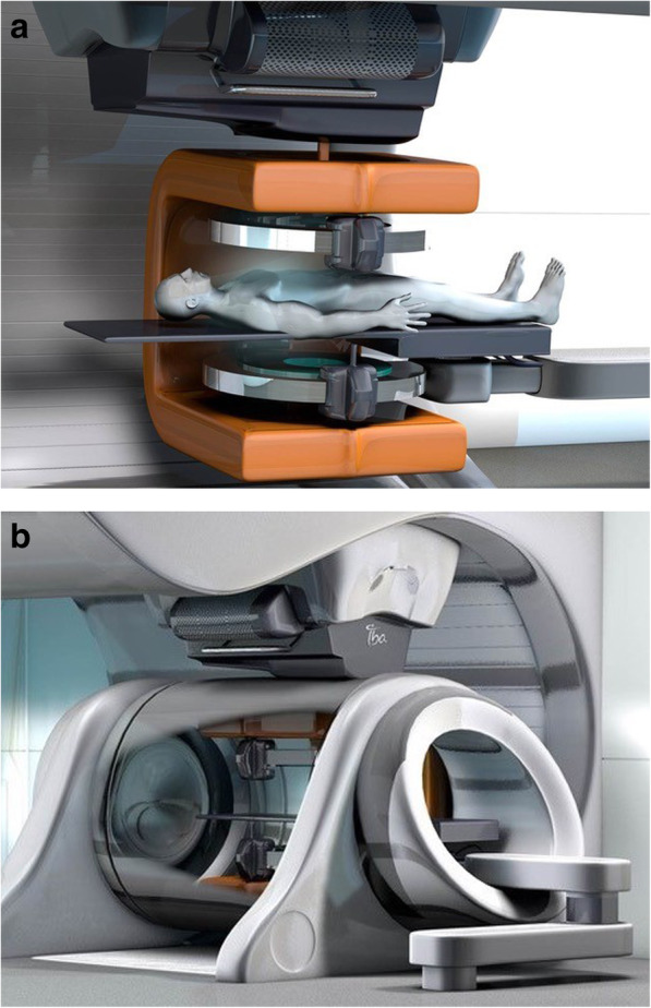
Background: The targeting accuracy of proton therapy (PT) for moving soft-tissue tumours is expected to greatly improve by real-time magnetic resonance imaging (MRI) guidance. The integration of MRI and PT at the treatment isocenter would offer the opportunity of combining the unparalleled soft-tissue contrast and real-time imaging capabilities of MRI with the most conformal dose distribution and best dose steering capability provided by modern PT. However, hybrid systems for MR-integrated PT (MRiPT) have not been realized so far due to a number of hitherto open technological challenges. In recent years, various research groups have started addressing these challenges and exploring the technical feasibility and clinical potential of MRiPT. The aim of this contribution is to review the different aspects of MRiPT, to report on the status quo and to identify important future research topics.
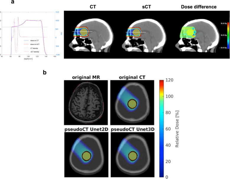
Methods: Four aspects currently under study and their future directions are discussed: modelling and experimental investigations of electromagnetic interactions between the MRI and PT systems, integration of MRiPT workflows in clinical facilities, proton dose calculation algorithms in magnetic fields, and MRI-only based proton treatment planning approaches.
Conclusions: Although MRiPT is still in its infancy, significant progress on all four aspects has been made, showing promising results that justify further efforts for research and development to be undertaken. First non-clinical research solutions have recently been realized and are being thoroughly characterized. The prospect that first prototype MRiPT systems for clinical use will likely exist within the next 5 to 10 years seems realistic, but requires significant work to be performed by collaborative efforts of research groups and industrial partners.
Keywords: Image guidance; Magnetic resonance imaging; Proton therapy.
Conflict of interest statement
The authors declare that they have no competing interests.
Figures


Similar articles
-
Toward MR-integrated proton therapy: modeling the potential benefits for liver tumors.Phys Med Biol. 2021 Sep 23;66(19). doi: 10.1088/1361-6560/ac1ef2. Phys Med Biol. 2021. PMID: 34407528
-
Technical note: Experimental dosimetric characterization of proton pencil beam distortion in a perpendicular magnetic field of an in-beam MR scanner.Med Phys. 2023 Nov;50(11):7294-7303. doi: 10.1002/mp.16448. Epub 2023 May 10. Med Phys. 2023. PMID: 37161832
-
Dosimetric feasibility of real-time MRI-guided proton therapy.Med Phys. 2014 Nov;41(11):111713. doi: 10.1118/1.4897570. Med Phys. 2014. PMID: 25370627 Free PMC article.
-
Magnetic resonance imaging (MRI) guided proton therapy: A review of the clinical challenges, potential benefits and pathway to implementation.Radiother Oncol. 2022 May;170:37-47. doi: 10.1016/j.radonc.2022.02.031. Epub 2022 Mar 5. Radiother Oncol. 2022. PMID: 35257848 Review.
-
Medical physics challenges in clinical MR-guided radiotherapy.Radiat Oncol. 2020 May 5;15(1):93. doi: 10.1186/s13014-020-01524-4. Radiat Oncol. 2020. PMID: 32370788 Free PMC article. Review.
Cited by
-
The history of ion beam therapy in Germany.Z Med Phys. 2022 Feb;32(1):6-22. doi: 10.1016/j.zemedi.2021.11.003. Epub 2022 Jan 31. Z Med Phys. 2022. PMID: 35101337 Free PMC article. Review.
-
Synthetic dual-energy CT for MRI-only based proton therapy treatment planning using label-GAN.Phys Med Biol. 2021 Mar 9;66(6):065014. doi: 10.1088/1361-6560/abe736. Phys Med Biol. 2021. PMID: 33596558 Free PMC article.
-
X-change symposium: status and future of modern radiation oncology-from technology to biology.Radiat Oncol. 2021 Feb 4;16(1):27. doi: 10.1186/s13014-021-01758-w. Radiat Oncol. 2021. PMID: 33541387 Free PMC article. Review.
-
Integrated MRI-guided radiotherapy - opportunities and challenges.Nat Rev Clin Oncol. 2022 Jul;19(7):458-470. doi: 10.1038/s41571-022-00631-3. Epub 2022 Apr 19. Nat Rev Clin Oncol. 2022. PMID: 35440773 Review.
-
SynthRAD2025 Grand Challenge dataset: Generating synthetic CTs for radiotherapy from head to abdomen.Med Phys. 2025 Jul;52(7):e17981. doi: 10.1002/mp.17981. Med Phys. 2025. PMID: 40665582 Free PMC article.
References
-
- MacKay RI. Image guidance for proton therapy. Clin Oncol (R Coll Radiol) 2018;30(5):293–298. - PubMed
-
- Landry G, Hua CH. Current state and future applications of radiological image guidance for particle therapy. Med Phys. 2018;45(11):e1086–e1095. - PubMed
-
- Corradini S, Alongi F, Andratschke N, Belka C, Boldrini L, Cellini F, Debus J, Guckenberger M, Hörner-Rieber J, Lagerwaard FJ, Mazzola R, Palacios MA, Philippens MEP, Raaijmakers CPJ, Terhaard CHJ, Valentini V, Niyazi M. MR-guidance in clinical reality: current treatment challenges and future perspectives. Radiat Oncol. 2019;14(1):92. - PMC - PubMed
-
- Oborn BM, Dowdell S, Metcalfe PE, Crozier S, Mohan R, Keall PJ. Future of medical physics: Real-time MRI-guided proton therapy. Med Phys. 2017;44(8):e77–e90. - PubMed
Publication types
MeSH terms
Grants and funding
LinkOut - more resources
Full Text Sources
Medical
