

Unusual presentation of a first Branchial cleft cyst associated with an abnormal bony canal -a case report
- PMID: 32471510
- PMCID: PMC7260795
- DOI: 10.1186/s40463-020-00426-5
Unusual presentation of a first Branchial cleft cyst associated with an abnormal bony canal -a case report
Abstract
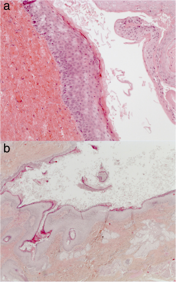
Background: First branchial cleft anomalies are rare, accounting for only 10% of all branchial cleft anomalies. We report an even more rare and unique case of a branchial cleft cyst with features of both first and second arch derivatives.
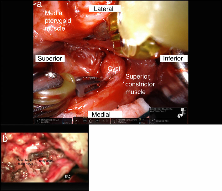
Case presentation: A 6-year-old boy presented to us with a left conductive hearing loss associated with pre-tympanic keratin debris and an ipsilateral painful cervical mass. He had a past medical history of left ear surgery for presumed cholesteatoma 2 years prior and left neck abscess drainage 6 months prior. CT and MRI revealed a lesion originating in the external auditory canal and extending cervically through a bony canal located medial to the facial nerve and terminating as a parapharyngeal cyst. The complete removal was accomplished in one surgical stage consisting of three distinct steps: robotic assisted transoral resection of the pharyngeal cyst, an endaural approach and a parotidectomy approach.
Conclusion: We believe that our detailed description of this rare first branchial cleft cyst with pharyngeal extension, possibly a hybrid case between a first and second branchial cyst, can serve as a valuable tool to Otolaryngologists - Head and Neck Surgeons who come across a similar unusual presentations.
Keywords: Cholesteatoma; Cyst; First branchial; Parapharyngeal; Robotic.
Conflict of interest statement
Not applicable.
Figures

Similar articles
-
First branchial cleft anomaly extending to parapharyngeal space.BMJ Case Rep. 2021 Aug 26;14(8):e244842. doi: 10.1136/bcr-2021-244842. BMJ Case Rep. 2021. PMID: 34446522 Free PMC article.
-
Bilateral Ear Canal Cholesteatoma with Underlying Type I First Branchial Cleft Anomalies.Ann Otol Rhinol Laryngol. 2019 Apr;128(4):360-364. doi: 10.1177/0003489418821700. Epub 2019 Jan 4. Ann Otol Rhinol Laryngol. 2019. PMID: 30607978 Free PMC article.
-
An unusual otoscopic finding associated with a type II first branchial cleft anomaly.J Laryngol Otol. 2012 Mar;126(3):316-8. doi: 10.1017/S0022215111003240. Epub 2011 Dec 13. J Laryngol Otol. 2012. PMID: 22152758
-
Duplicated facial nerve trunk with a first branchial cleft cyst.Laryngoscope. 2014 Mar;124(3):662-4. doi: 10.1002/lary.24365. Epub 2013 Sep 19. Laryngoscope. 2014. PMID: 23946158 Review.
-
A unique location of branchial cleft cyst: case report and review of the literature.Int J Oral Maxillofac Surg. 2019 Jun;48(6):712-715. doi: 10.1016/j.ijom.2018.11.014. Epub 2018 Dec 20. Int J Oral Maxillofac Surg. 2019. PMID: 30579743 Review.
Cited by
-
Current Applications and Outcomes of Robotic Surgery in Pediatric Upper Airway and Neck Procedures: A Systematic Review.Children (Basel). 2025 Jun 13;12(6):765. doi: 10.3390/children12060765. Children (Basel). 2025. PMID: 40564723 Free PMC article. Review.
-
Unusual presentation of a first branchial arch fistula with maxillofacial infection: a case report.BMC Surg. 2021 Jul 3;21(1):306. doi: 10.1186/s12893-021-01303-2. BMC Surg. 2021. PMID: 34217239 Free PMC article.
-
Isolated First Branchial Cleft Anomalies of the External Auditory Canal.Am J Case Rep. 2022 Nov 7;23:e936809. doi: 10.12659/AJCR.936809. Am J Case Rep. 2022. PMID: 36342864 Free PMC article.
-
First branchial cleft anomaly extending to parapharyngeal space.BMJ Case Rep. 2021 Aug 26;14(8):e244842. doi: 10.1136/bcr-2021-244842. BMJ Case Rep. 2021. PMID: 34446522 Free PMC article.
References
-
- Chandler J, Mitchell B. Branchial cleft cysts, sinuses and fistulas. Otolaryngol Clin N Am. 1981;14(1):175–186. - PubMed
-
- Work W. Newer concepts of first branchial cleft defects. Laryngoscope. 1972;82(9):1581–1593. - PubMed
-
- Gupta A-K, Kumar S, Jain A. Bilateral first and second branchial cleft fistulas: a case report. Ear Nose Throat J. 2008;87(5):291–293. - PubMed
-
- Castallenos A, Scangas GA, Naunheim MR, Raol N, Cohen MS. Avoiding surgical pitfalls during resection of a “hybrid” first and second branchial cleft cyst- a case report. Int J Pediatr Otorhinolaryngol. 2016;87:91–93. - PubMed
Publication types
MeSH terms
LinkOut - more resources
Full Text Sources
Medical
Miscellaneous
