. 2020 Jul 20;86(15):e01026-20.
doi: 10.1128/AEM.01026-20. Print 2020 Jul 20.

Specific Root Exudate Compounds Sensed by Dedicated Chemoreceptors Shape Azospirillum brasilense Chemotaxis in the Rhizosphere

Affiliations

Specific Root Exudate Compounds Sensed by Dedicated Chemoreceptors Shape Azospirillum brasilense Chemotaxis in the Rhizosphere

Lindsey O'Neal et al. Appl Environ Microbiol. .

Abstract

Plant roots shape the rhizosphere community by secreting compounds that recruit diverse bacteria. Colonization of various plant roots by the motile alphaproteobacterium Azospirillum brasilense causes increased plant growth, root volume, and crop yield. Bacterial chemotaxis in this and other motile soil bacteria is critical for competitive colonization of the root surfaces. The role of chemotaxis in root surface colonization has previously been established by endpoint analyses of bacterial colonization levels detected a few hours to days after inoculation. More recently, microfluidic devices have been used to study plant-microbe interactions, but these devices are size limited. Here, we use a novel slide-in chamber that allows real-time monitoring of plant-microbe interactions using agriculturally relevant seedlings to characterize how bacterial chemotaxis mediates plant root surface colonization during the association of A. brasilense with Triticum aestivum (wheat) and Medicago sativa (alfalfa) seedlings. We track A. brasilense accumulation in the rhizosphere and on the root surfaces of wheat and alfalfa. A. brasilense motile cells display distinct chemotaxis behaviors in different regions of the roots, including attractant and repellent responses that ultimately drive surface colonization patterns. We also combine these observations with real-time analyses of behaviors of wild-type and mutant strains to link chemotaxis responses to distinct chemicals identified in root exudates to specific chemoreceptors that together explain the chemotactic response of motile cells in different regions of the roots. Furthermore, the bacterial second messenger c-di-GMP modulates these chemotaxis responses. Together, these findings illustrate dynamic bacterial chemotaxis responses to rhizosphere gradients that guide root surface colonization.IMPORTANCE Plant root exudates play critical roles in shaping rhizosphere microbial communities, and the ability of motile bacteria to respond to these gradients mediates competitive colonization of root surfaces. Root exudates are complex chemical mixtures that are spatially and temporally dynamic. Identifying the exact chemical(s) that mediates the recruitment of soil bacteria to specific regions of the roots is thus challenging. Here, we connect patterns of bacterial chemotaxis responses and sensing by chemoreceptors to chemicals found in root exudate gradients and identify key chemical signals that shape root surface colonization in different plants and regions of the roots.

Keywords: Azospirillum; chemotaxis; rhizosphere; root exudates.


Figures

FIG 1
FIG 1
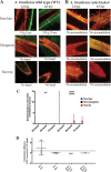
Interaction of WT and nonchemotactic (Δche1 Δche4) A. brasilense with wheat roots in the slide-in chamber. WT and chemotaxis-null (Δche1 Δche4) bacteria carrying pHRGFP to constitutively express GFP were mixed with the semisolid molten medium used to fill the chamber. Seedlings were planted at the top of the chamber (see Materials and Methods for details). Bacteria are visible in green, and the wheat roots are in red. Shown are WT (A) and Δche1 Δche4 (B) accumulations close to the root surface in the root hair and elongation zones. Bands of motile fluorescent bacteria are denoted by brackets, and the average bandwidths in micrometers are noted below the pictures. Images are representative of data from 3 biological replicates in different chambers.
FIG 2
FIG 2
Quantification of A. brasilense colonization levels on wheat roots. Colonization levels in each root zone were determined by measuring the fluorescence of surface-attached cells normalized to the area of the root observed and expressed in arbitrary units (a.u.). The calculated total corrected fluorescence (CTCF) for each channel was calculated as follows: integrated density/(area of selection × mean fluorescence of the background). t tests were used to determine if WT colonization differed significantly from Δche1 Δche4 colonization. * indicates a significant difference from the Δche1 Δche4 strain (P ≤ 0.05).
FIG 3
FIG 3
WT and chemotaxis-null (Δche1 Δche4) behaviors in the presence of alfalfa roots. (A and B) Free-swimming WT (A) and Δche1Δche4 (B) cell accumulation around the alfalfa roots in the slide-in chamber. Shown is the time course of colonization of alfalfa roots by the WT (A) and Δche1 Δche4 (B) strains in the slide-in chamber. A. brasilense bacteria constitutively expressing GFP were inoculated into a slide-in chamber containing 3-day-old wheat and imaged over the course of 72 h postinoculation (hpi). A. brasilense is visible in green, and the wheat roots are in red (autofluorescence). Images are representative of results from 3 chamber replicates. (C) In-chamber colonization levels were quantified by calculating the CTCF of the attached bacteria and normalizing to the root area. (D) Five-day colonization was measured by CFU counts from homogenized roots. A t test indicated no significant difference from Δche1 Δche4 colonization (P ≤ 0.05) for colonization in any of the root zones. n.r., no bacteria recovered.
FIG 4
FIG 4
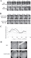
Interaction of wild-type and nonchemotactic (Δche1 Δche4) A. brasilense bacteria in the root-in-pool assay with intact roots from wheat and alfalfa seedlings. (A and B) Wheat roots from intact seedlings were placed in a suspension of motile WT (A) or chemotaxis-null mutant (B) strain bacteria, and free-swimming behavior in the vicinity of the roots was observed and recorded over time. WT A. brasilense accumulates in a band in the root hair and tip zones (indicated by the white arrows), while the chemotaxis-null strain forms a homogeneous pool that does not change within 10 min of observation. (C) Intensity profile analysis of images shown for the WT cell suspension in the root-in-pool assay at 0, 30, and 60 s. (D) WT and chemotaxis-null strains in the presence of the root tip (left) and root hair zone (right) of alfalfa seedlings in the root-in-pool assay.
FIG 5
FIG 5
Principal-component analysis (PCA) of metabolites detected in wheat and alfalfa root exudates. (A) PCA loading plot of the log10 abundance of total metabolites isolated from three biological samples of wheat and alfalfa. The 95% confidence intervals of the PCA scores’ covariances from three samples of wheat and alfalfa are represented as ellipses. (B) PCA of only organic acids isolated from three biological samples of wheat and alfalfa. The 95% confidence intervals of the PCA scores’ covariances from 3 samples of wheat and alfalfa are represented as ellipses. (C) PCA loading plot of log10 abundances of only amino acids isolated from three biological samples of wheat and alfalfa. The 95% confidence intervals of the PCA scores’ covariances from 3 samples of wheat and alfalfa are represented as ellipses.
FIG 6
FIG 6
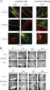
A. brasilense Δtlp1 mutant strain free-swimming response and colonization of wheat roots. (A) Δtlp1 strain in the slide-in chamber with wheat seedlings. Shown is the time course of the colonization of wheat root surfaces by the A. brasilense Δtlp1 mutant strain. The chambers were filled with Fahraeus medium containing A. brasilense constitutively expressing GFP (pHRGFP) and imaged over the course of 72 h. A. brasilense is visible in green, and the wheat roots are in red. Images are representative of data from 3 biological replicates in different chambers. Colonization levels were quantified by calculating the CTCF for each channel as follows: integrated density/(area of selection × mean fluorescence of the background). A t test indicated no significant difference from WT A. brasilense colonization (P ≤ 0.05) for colonization in any of the root zones. (B) Role of Tlp1 and c-di-GMP binding to Tlp1 in the response to wheat roots. A root-in-pool assay was used to observe the behavior of cells lacking Tlp1 (Δtlp1) or expressing Tlp1 impaired in binding to c-di-GMP (Tlp1R562A R563A) in the presence of wheat. Arrows indicate the accumulation of motile cells at that position.
FIG 7
FIG 7
Roles of PilZ domain chemoreceptors and c-di-GMP levels in the response to ROS gradients. (A and B) Motile A. brasilense Δtlp1 (A) or Δaer (B) bacteria were exposed to gradients generated by filter paper soaked in buffer, hydrogen peroxide, or cumene peroxide. (C and D) To determine the role of intracellular c-di-GMP levels in the response to ROS gradients, c-di-GMP levels were manipulated using an optogenetic plasmid system, as described in Materials and Methods. WT A. brasilense cells with a red-light-activated diguanylate cyclase (pRED-DGC) or blue-light-activated phosphodiesterase (pBLUE-PDE) were illuminated with green (control), red, or blue light before exposure to filter paper soaked in Che buffer or various concentrations of hydrogen peroxide. A chemotactic response is indicated by a ring forming a certain distance away from the filter paper. Black arrows indicate an accumulation of motile cells. Positive responses are denoted by black arrows pointing at the ring formed by bacteria. The plates were observed every 5 min.

Similar articles

Cited by

References

    1. Souza RD, Ambrosini A, Passaglia LMP. 2015. Plant growth-promoting bacteria as inoculants in agricultural soils. Genet Mol Biol 38:401–419. doi:10.1590/S1415-475738420150053. - DOI - PMC - PubMed
    1. Glick BR. 2012. Plant growth-promoting bacteria: mechanisms and applications. Scientifica (Cairo) 2012:963401. doi:10.6064/2012/963401. - DOI - PMC - PubMed
    1. Philippot L, Raaijmakers JM, Lemanceau P, van der Putten WH. 2013. Going back to the roots: the microbial ecology of the rhizosphere. Nat Rev Microbiol 11:789–799. doi:10.1038/nrmicro3109. - DOI - PubMed
    1. Hinsinger P, Bengough AG, Vetterlein D, Young IM. 2009. Rhizosphere: biophysics, biogeochemistry and ecological relevance. Plant Soil 321:117–152. doi:10.1007/s11104-008-9885-9. - DOI
    1. Massalha H, Korenblum E, Malitsky S, Shapiro OH, Aharoni A. 2017. Live imaging of root-bacteria interactions in a microfluidics setup. Proc Natl Acad Sci U S A 114:4549–4554. doi:10.1073/pnas.1618584114. - DOI - PMC - PubMed

Publication types

LinkOut - more resources