. 2020 May 29;11(5):407.
doi: 10.1038/s41419-020-2622-x.

Crosstalk between androgen and Wnt/β-catenin leads to changes of wool density in FGF5-knockout sheep

Affiliations

Crosstalk between androgen and Wnt/β-catenin leads to changes of wool density in FGF5-knockout sheep

Rui Zhang et al. Cell Death Dis. .

Abstract

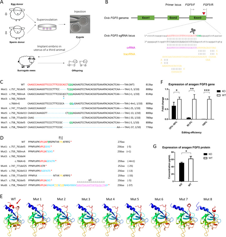
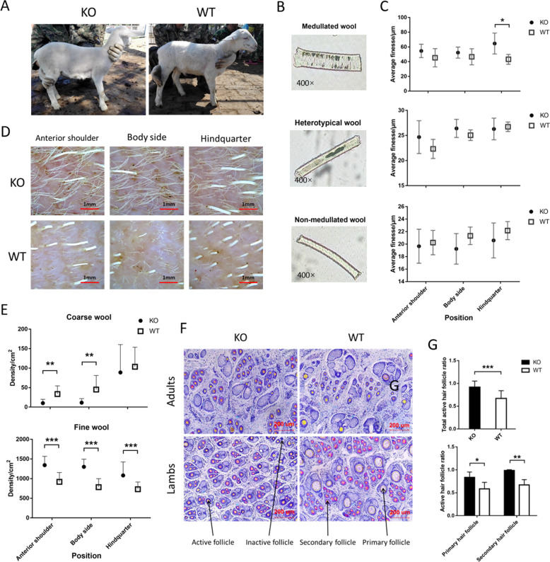
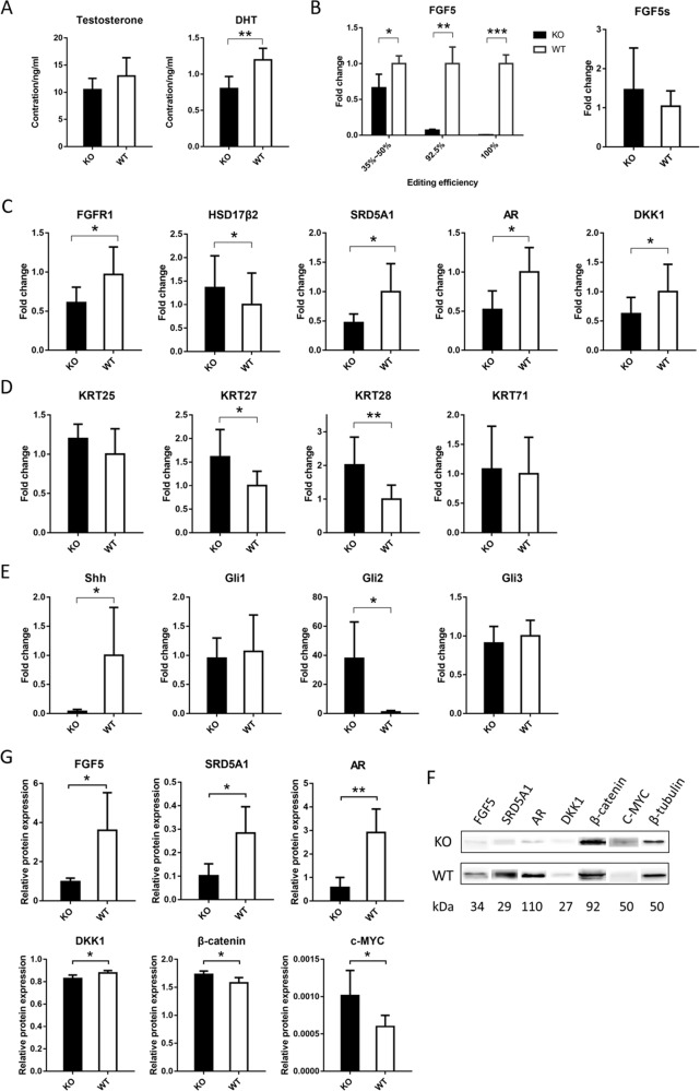
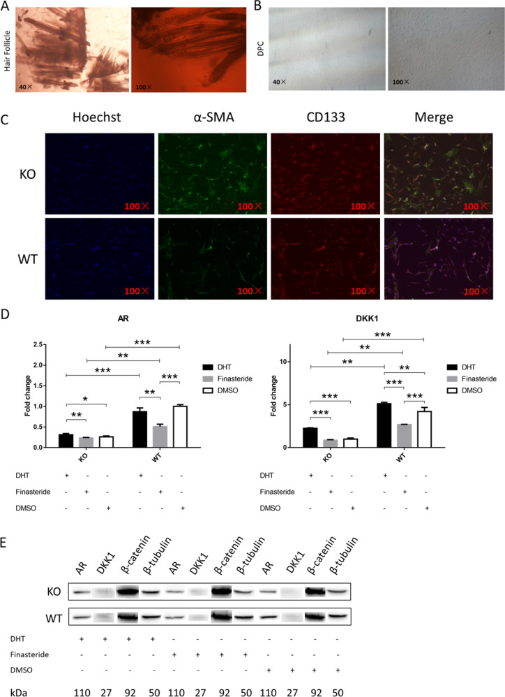
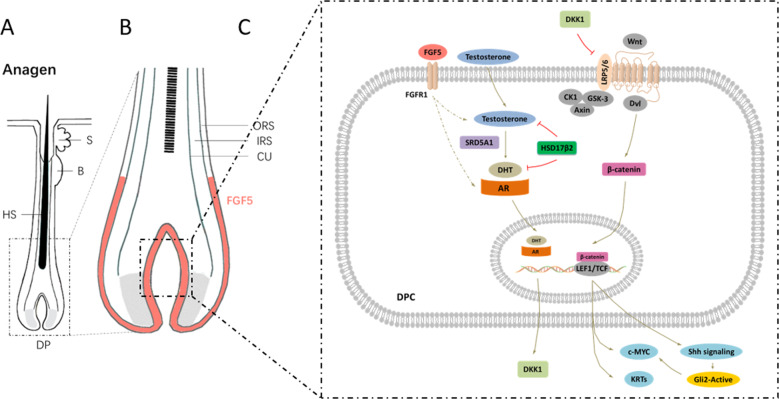
Fibroblast growth factor 5 (FGF5) is a famous dominant inhibitor of anagen phase of hair cycle. Mutations of FGF5 gene result in a longer wool in mice, donkeys, dogs, cats, and even in human eyelashes. Sheep is an important source of wool production. How to improve the production of wool quickly and effectively is an urgent problem to be solved. In this study, we generated five FGF5-knockout Dorper sheep by the CRISPR/Cas9 system. The expression level of FGF5 mRNA in knockout (KO) sheep decreased significantly, and all FGF5 proteins were dysfunctional. The KO sheep displayed a significant increase in fine-wool and active hair-follicle density. The crosstalk between androgen and Wnt/β-catenin signaling downstream of FGF5 gene plays a key role. We established downstream signaling cascades for the first time, including FGF5, FGFR1, androgen, AR, Wnt/β-catenin, Shh/Gli2, c-MYC, and KRTs. These findings further improved the function of FGF5 gene, and provided therapeutic ideas for androgen alopecia.


Conflict of interest statement

The authors declare that they have no conflict of interest.

Figures

Fig. 1
Fig. 1. Generation and screening of FGF5-knockout sheep.
a Representative schematic of the experimental design. After mating and superovulation of the donor sheep, Cas9 mRNA and sgRNA were co-injected into one-cell embryos; then the embryo was implanted into the uterus of a third animal. The editing efficiencies were detected after the birth of the lambs. b The targeted sequence and the detecting primer sequences at sheep FGF5 locus. Red triangle indicates the predicted DSB cleavage site for the sgRNA. The PAM and protospacer sequences are highlighted in green and red, respectively. c Schematic diagram of the modified FGF5 partial protein-coding region and the targeting locus of sgRNA: Cas9. sgRNA-targeting sites are presented in red text; PAM sequences are highlighted in green and underlined; the mutations are blue, lower case; insertions (+), deletions (−), mutation (m), and the frequencies occurring in individuals are shown to the right of each allele. d Schematic diagram of the changes in partial protein AA sequences of modified FGF5 in KO sheep. Protein AA sequences of the sgRNA-targeting site are presented in red text; protein AA sequences of PAM sequence are highlighted in green and underlined; the deletions and changes in protein AA sequences are highlighted in blue; the secondary structure of β-strands is highlighted in yellow, and changes in β-strands caused by mutations are underlined; insertions (+), deletions (−), and mutation (m) are shown to the right of each allele. e Changes in the tertiary structure of different mutants. The mutated FGF5 lacked the β12 strands (marked in dark-red arrows). f The RNA expression of FGF5 decreases in KO individuals with different editing efficiencies in anagen. g The protein expression of FGF5 in the KO group also decreases compared with the control group in anagen by ELISA.
Fig. 2
Fig. 2. Changes in sheep wool and hair-follicle density after FGF5 gene editing in anagen.
a Photographs of one of the KO sheep and its sibling. b There are three types of wool in the KO and the control group. c The changes in fineness of three types of wool in three different parts of KO and control group. Except for the medullated wool of the KO group at the hindquarter, the fineness of which was significantly higher than the control group, there was no significant difference in the fineness of the wools in the other parts. d Photograph of skin follicles at 48 h after shaving. The actual area shown in the picture is 16 mm². e Changes in coarse- and fine-wool density in the three parts of the KO and control group. The densities of fine wool in the KO group were significantly higher than that in the control group at all three sites. f Sacpic staining of adult sheep and lamb skin sections in the KO and control group. The ruler is marked in red in the lower-right corner of the picture. Primary, secondary, active, and inactive hair follicles are also labeled with arrows, respectively. g Changes in the total active hair-follicle ratio of coarse and fine wool between the KO and control group. Regardless of the total active hair-follicle ratio or the proportion of active primary and secondary hair follicles, the KO group was significantly higher than the control group. The statistical results in panel e are from d, and the statistical results in panel g are from f.
Fig. 3
Fig. 3. FGF5-mediated multiple signaling promotes the increase in wool density and the active hair-follicle density in anagen.
a Changes in testosterone and DHT in the skin tissue of KO and control groups. The expression level of DHT in the skin tissue of the KO group was significantly lower than that of the control group. b Changes in FGF5 in the skin tissue of the KO and control group. The expression levels of FGF5 gene in different editing efficiency individuals were different, but they were significantly lower than the control group. However, the expression level of the FGF5s gene did not change significantly. c Expression changes of the related genes in AR and Wnt signaling in the skin tissue of the KO and control group. d Expression changes of IRS-related KRTs in the skin tissue of the KO and control group. e Expression changes of Shh signaling in the skin tissue of the KO and control group. f The expression levels of FGF5, SRD5A1, AR, DKK1, β-catenin, and c-MYC proteins in the skin tissue were detected by western blotting. The amount of β-tubulin was used as a control. g Compared with the control group, the protein expression levels of FGF5, SRD5A1, AR, DKK1, β-catenin, and c-MYC in the KO group were significantly changed.
Fig. 4
Fig. 4. Addition of DHT and finasteride to DPCs demonstrates the interaction between androgen and Wnt/β-catenin.
a Hair follicles of the KO and control group are obtained from skin tissue. b Dermal papilla cells are isolated and purified with a cloning loop. c The isolated dermal papilla cells were subjected to immunofluorescence staining. The positive α-SMA proved to be a dermal papilla cell, and CD133- positive proved that the cell was in the growth phase. d Changes in the relative expression levels of AR and DKK1 genes in DPCs of KO and the control group after the addition of DHT, finasteride, and DMSO, respectively. e Changes in the relative protein levels of AR and β-catenin in DPCs of KO and the control group after the addition of DHT, finasteride, and DMSO, respectively.
Fig. 5
Fig. 5. Diagram illustrating the signaling pathways involved in FGF5 gene-editing sheep during anagen.
a Schematic diagram of hair follicles during anagen. b An enlarged view of the DP area and the signaling pathways that occur in it. c The molecular mechanism of FGF5 signaling cascade occurs in DPC, which changes the wool and the active hair-follicle density. The FGF5 protein secreted by the outer root sheath cells binds to its receptor FGFR1, causing changes in related signals in DPCs. Among them, the expression levels of SRD5A1 and HSD17β2 were changed, and the process of testosterone to DHT was also regulated. DHT can bind to AR and further into the nucleus, which positively regulates the expression of genes such as DKK1. As an inhibitor of the Wnt signaling pathway, DKK1 cascades the AR signaling pathway, and the Wnt/β-catenin signaling pathway to regulate the growth and development of hair follicles. In addition, the Shh signaling pathway and IRS-related KRTs and C-MYC also participate in the whole process as part of the downstream of the Wnt/β-catenin signaling pathway.

Similar articles

Cited by

References

    1. Stenn KS, Paus R. Controls of hair follicle cycling. Physiol. Rev. 2001;81:449. - PubMed
    1. Schneider MR, Schmidt-Ullrich R, Paus R. The hair follicle as a dynamic miniorgan. Curr. Biol. 2009;19:R132. - PubMed
    1. Krause K, Foitzik K. Biology of the hair follicle: the basics. Semin. Cutan. Med. Surg. 2006;25:2. - PubMed
    1. Fuchs, E., Merrill, B. J., Jamora, C. & DasGupta, R. At the Roots of a Never-Ending Cycle. Dev. Cell1, 13–25 (2001). - PubMed
    1. Beenken A, Mohammadi M. The FGF family: biology, pathophysiology and therapy. Nat. Rev. Drug. Discov. 2009;8:235. - PMC - PubMed

Publication types

MeSH terms