. 2020 May 29;3(1):273.
doi: 10.1038/s42003-020-0979-0.

A mouse model that is immunologically tolerant to reporter and modifier proteins

Affiliations

A mouse model that is immunologically tolerant to reporter and modifier proteins

Kaspar Bresser et al. Commun Biol. .

Abstract

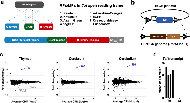
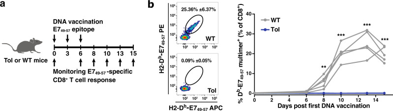
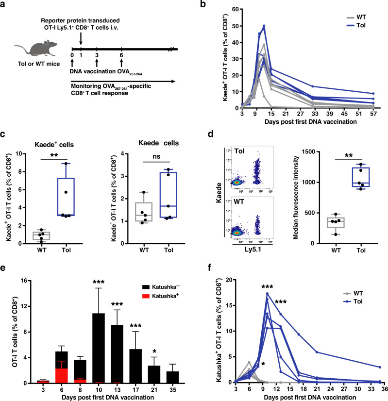
Reporter proteins have become an indispensable tool in biomedical research. However, exogenous introduction of these reporters into mice poses a risk of rejection by the immune system. Here, we describe the generation, validation and application of a multiple reporter protein tolerant 'Tol' mouse model that constitutively expresses an assembly of shuffled reporter proteins from a single open reading frame. We demonstrate that expression of the Tol transgene results in the deletion of CD8+ T cells specific for a model epitope, and substantially improves engraftment of reporter-gene transduced T cells. The Tol strain provides a valuable mouse model for cell transfer and viral-mediated gene transfer studies, and serves as a methodological example for the generation of poly-tolerant mouse strains.


Conflict of interest statement

The authors declare no competing interests.

Figures

Fig. 1
Fig. 1. Generation and validation of the Tol mouse model.
a Top left: cartoon depicting the strategy for reporter gene fragmentation, applied for each RP/MP gene included in the Tol ORF. Top right: reporter proteins included in the Tol ORF. Bottom: schematic overview of gene fragment placement in the Tol ORF. Note that the Tol ORF encodes the HPV E749–57 epitope in the COOH-terminal region of the chimeric Tol protein. b Cartoon depicting the targeting strategy of the Tol ORF into the Col1a1 locus through recombinase-mediated cassette exchange. c Differential gene expression analysis of indicated organs of Tol mice relative to WT mice. Scatterplots indicate average log2 fold changes and average counts per million. Genes with a statistically significant change in expression in Tol mice are indicated in gray, and the Tol transcript is indicated in blue. d Expression levels of the Tol transcript in thymi of Tol and WT mice. Data shown (c, d) is aggregated from n = 4 (Tol) and n = 4 (WT) mice. RP reporter protein, MP modifier protein, RMCE recombinase-mediated cassette exchange, PURO-R puromycin resistance gene, TK thymidine kinase gene, CPM counts per million.
Fig. 2
Fig. 2. Complete loss of E749–57 immunogenicity in Tol mice.
a Schematic overview of vaccination and monitoring strategy. b Left: Representative flow cytometry plots depicting E749–57-specific CD8+ T cells at day 13 post DNA vaccination in WT and Tol mice. Right: E749–57-specific CD8+ T-cell response in blood of WT (gray) and Tol (blue) mice at the indicated timepoints post first DNA vaccination. Lines indicate individual mice. Cells are gated on IR-dyeCD8+ lymphocytes. Depicted data are representative of two independent experiments, including at least 5 mice per group. P values were determined by Repeated Measures two-way ANOVA with Sidak’s multiple comparisons test to test the significance at each timepoint. *p < 0.05; **p < 0.01; ***p < 0.001.
Fig. 3
Fig. 3. Enhanced engraftment of Kaede- and Katushka-expressing cells in Tol mice.
a Schematic overview of experimental design. b Percentage of Kaede+ OT-I T cells in the blood of WT (gray) and Tol (blue) mice at the indicated timepoints post first DNA vaccination. c Percentage of Kaede+ (left) or Kaede (right) OT-I cells in WT and Tol mice at day 57 after first DNA vaccination. d Left: representative flow cytometry plots depicting Kaede expression in transferred (Ly5.1+) cells at day 57 after first DNA vaccination. Right: median fluorescence intensity (MFI) of Kaede+ OT-I cells in WT and Tol mice at day 57 post first DNA vaccination. e Percentage of Katushka+ (red) and Katushka (black) OT-I cells in WT animals at the indicated timepoints after first DNA vaccination. f Percentage Katushka+ OT-I cells in WT and Tol mice at the indicated timepoints after first DNA vaccination. Cells (bf) are gated on IR-dyeCD8+ lymphocytes, and plots depict n = 5 (Tol) and n = 5 (WT) mice. Depicted data are representative of one (bd) or two (e, f) independent experiments. Box-plots (c, d) depict interquartile range, with whiskers representing minimum and maximum values. Dots (c, d) and lines (b, f) indicate individual mice. Error bars (e) represent SD. P values were determined by either two-tailed Mann–Whitney signed rank test (c, d) or Repeated Measures two-way ANOVA with Sidak’s multiple comparisons test (e, f). *p < 0.05; **p < 0.01; ***p < 0.001, ns not significant.

References

    1. Ghim C-M, Lee SK, Takayama S, Mitchell RJ. The art of reporter proteins in science: past, present and future applications. BMB Rep. 2010;43:451–460. doi: 10.5483/BMBRep.2010.43.7.451. - DOI - PubMed
    1. Chalfie M, Tu Y, Euskirchen G, Ward WW, Prasher DC. Green fluorescent protein as a marker for gene expression. Science. 1994;263:802–805. doi: 10.1126/science.8303295. - DOI - PubMed
    1. Shaner NC, et al. Improved monomeric red, orange and yellow fluorescent proteins derived from Discosoma sp. red fluorescent protein. Nat. Biotechnol. 2004;22:1567–1572. doi: 10.1038/nbt1037. - DOI - PubMed
    1. Orban PC, Chui D, Marth JD. Tissue- and site-specific DNA recombination in transgenic mice. PNAS. 1992;89:6861–6865. doi: 10.1073/pnas.89.15.6861. - DOI - PMC - PubMed
    1. Han WGH, Unger WWJ, Wauben MHM. Identification of the immunodominant CTL epitope of EGFP in C57BL/6 mice. Gene Ther. 2008;15:700–701. doi: 10.1038/sj.gt.3303104. - DOI - PubMed

Publication types