Review
. 2020 Sep;20(9):533-549.
doi: 10.1038/s41568-020-0265-y. Epub 2020 May 29.

Tumour predisposition and cancer syndromes as models to study gene-environment interactions

Affiliations
Review

Tumour predisposition and cancer syndromes as models to study gene-environment interactions

Michele Carbone et al. Nat Rev Cancer. 2020 Sep.

Abstract

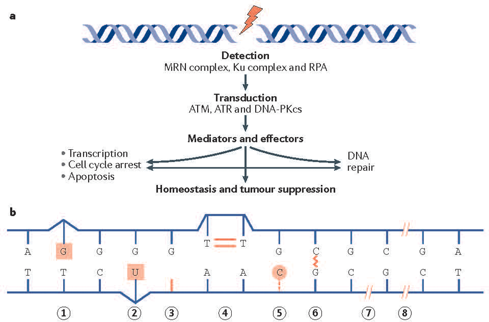
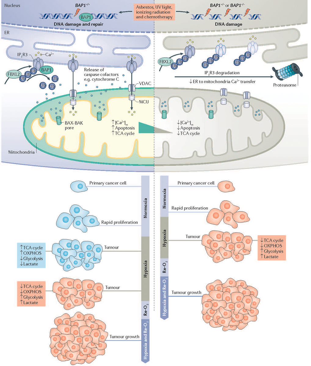
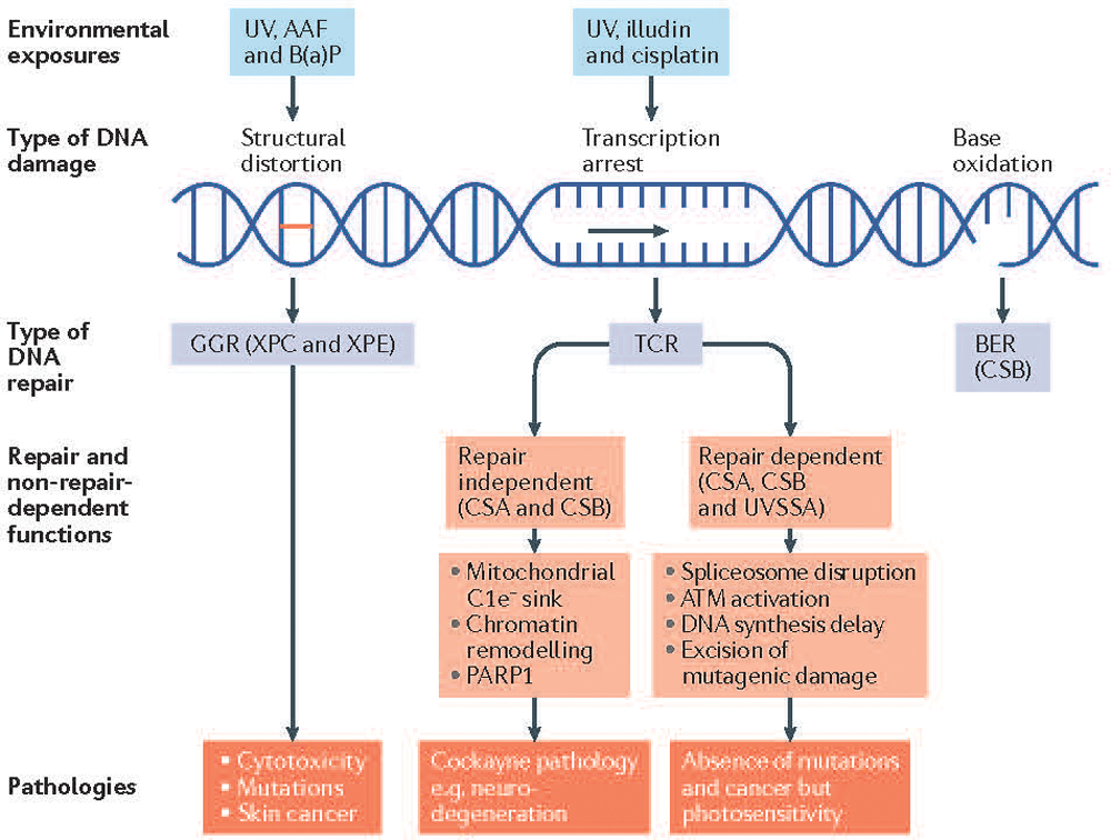
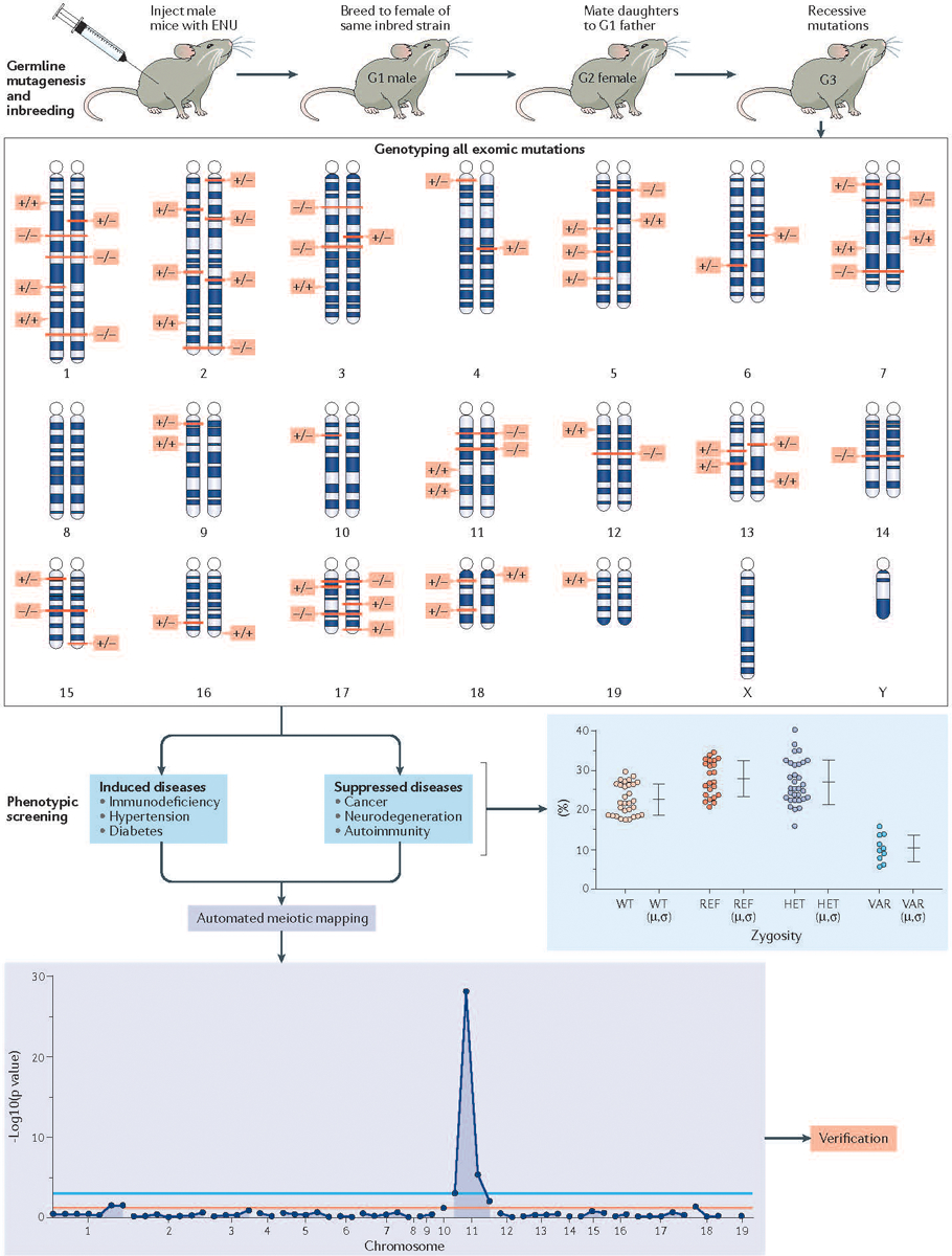
Cell division and organismal development are exquisitely orchestrated and regulated processes. The dysregulation of the molecular mechanisms underlying these processes may cause cancer, a consequence of cell-intrinsic and/or cell-extrinsic events. Cellular DNA can be damaged by spontaneous hydrolysis, reactive oxygen species, aberrant cellular metabolism or other perturbations that cause DNA damage. Moreover, several environmental factors may damage the DNA, alter cellular metabolism or affect the ability of cells to interact with their microenvironment. While some environmental factors are well established as carcinogens, there remains a large knowledge gap of others owing to the difficulty in identifying them because of the typically long interval between carcinogen exposure and cancer diagnosis. DNA damage increases in cells harbouring mutations that impair their ability to correctly repair the DNA. Tumour predisposition syndromes in which cancers arise at an accelerated rate and in different organs - the equivalent of a sensitized background - provide a unique opportunity to examine how gene-environment interactions influence cancer risk when the initiating genetic defect responsible for malignancy is known. Understanding the molecular processes that are altered by specific germline mutations, environmental exposures and related mechanisms that promote cancer will allow the design of novel and effective preventive and therapeutic strategies.


Conflict of interest statement

Competing interests

S.T.A., B.B., A.B., W.C., J.E.C., C.M.C., W.D.F., G.G., J.L.G., E.P.H., P.M.H., T.W.M., D.M., F.N., declare no competing interests.

Figures

Figure 1.
Figure 1.. DNA repair pathways and cancer.
(A) The DNA damage response is activated by DNA damage sensors that include the MRE11–RAD50–NBS1 (MRN) complex and the Ku heterodimer, which detect DNA double-strand breaks. The replication protein A (RPA) heterotrimer engages exposed regions of single stranded DNA. These sensors recruit the transducing kinases ataxia telangiectasia mutated (ATM) (through the MRN complex), DNA-dependent protein kinase catalytic subunit (DNA-PKcs) (through the Ku heterodimer), and ataxia telangiectasia and Rad3-related protein (ATR) (through RPA), which in turn phosphorylate a broad spectrum of mediators and effectors. Ultimately the DNA damage response promotes survival at the cellular level and homeostasis and tumor suppression at the organismal level. (B) Depicted are DNA lesions that can promote cancer development. Mismatch repair (MMR) corrects mismatched base pairs (lesion 1). Base excision repair [G] (BER) removes chemically damaged bases (lesion 2), repairs abasic sites (a location in DNA that has neither a purine nor a pyrimidine base due to DNA damage) (lesion 3) and repairs DNA nicks (where there is no phosphodiester bond between adjacent nucleotides of one strand) (lesion 7). Nucleotide excision repair (NER) is required for the removal of cyclobutane pyrimidine dimers (lesion 4) and 6–4 photoproducts (not shown) arising from exposure to ultraviolet (UV) light. NER and BER, along with alkyl transferases are responsible for removal of damaged bases and bulky adducts (lesion 5). The Fanconi anemia (FA) pathway removes intra-strand cross links (lesion 6). DNA double strand breaks can cause pathogenic DNA rearrangements, and are repaired by non-homologous end joining [G] (NHEJ) and homologous recombination (HR) pathways (lesion 8). The reader is directed to a comprehensive review.
Figure 2.
Figure 2.. Mechanisms of BAP1 activity in cancer development.
The powerful tumor suppressor activity of BRCA1-associated protein 1 (BAP1) and its ability to regulate gene x environment interactions (GxE) in carcinogenesis are related to its dual role in the nucleus, where BAP1 contributes to DNA repair by modulating homologous recombination (HR), and in the cytoplasm where BAP1 regulates cell death and mitochondrial respiration. In the cytoplasm, BAP1 localizes at the endoplasmic reticulum (ER) where it binds, deubiquitylates (following F-box and leucine-rich repeat protein 2 (FBXL2) ubiquitylation), and stabilizes type 3 inositol-1,4,5-trisphosphate receptor (IP3R3), modulating calcium ion (Ca2+) release from the ER into the cytosol and mitochondria, and thus promoting apoptosis. In primary cells exposed to either asbestos, ionizing radiation (IR) or ultraviolet (UV) radiation, reduced levels of nuclear BAP1 impair DNA repair. At the same time, reduced cytoplasmic BAP1 levels impair apoptosis, increasing the fraction of cells that survive DNA damage and that over time may become malignant. In addition, mitochondria need Ca2+ for aerobic oxidative phosphorylation (OXPHOS). In response to tumor hypoxia, cancer cells need to adjust their metabolism from aerobic (blue) to glycolytic (red) in order to sustain growth and survival. Primary cells from BAP1+/− individuals have reduced mitochondrial OXPHOS and increased aerobic glycolysis and lactate production, even in the presence of oxygen, a phenomenon known as the ‘Warburg effect’. Therefore, the ‘Warburg effect’ in addition to being a hallmark of cancer cells is also found in normal cells from BAP1-mutant carriers, and contributes to the adaptation to metabolic stress during tumorigenesis. BAP1+/+, cells with BAP1 wild-type; BAP1+/−, cells with heterozygous BAP1 mutations, containing about 50% of BAP1 protein levels compared to wild-type cells. [Ca2+]m, mitochondrial calcium; MCU, mitochondrial calcium uniporter; Re-O2 reoxygenation; TCA, tricarboxylic acid; Ub, ubiquitin; VDAC, voltage-dependent anion channel.
Figure 3.
Figure 3.. Xeroderma pigmentosum and Cockayne syndrome as examples of environmental impacts and genetics on DNA damage and repair.
Pathways activated by DNA damage depend on the nature of the damage, cell cycle stage, and the DNA damage response. The proteins in these pathways also have roles beyond DNA repair. DNA damage from exogenous agents that distort the DNA double helix creates a target for global genome repair (GGR) also known as GG-nucleotide excision repair (GG-NER). Examples include N-2-acetylaminofluorene (AAF) (a carcinogenic and mutagenic derivative of fluorene that forms adducts at the C8 position of guanine in DNA) and benzo[a]pyrene (B(a)P) (a polycyclic aromatic hydrocarbon that also forms DNA adducts). Xeroderma pigmentosum (XP), mutations in the damage recognition proteins xeroderma pigmentosum complementation group E (XPE) and XPC cause increased ultraviolet (UV)-specific mutations and cancer in exposed tissues, mainly skin. Agents such as the fungal toxin illudin and the chemotherapeutic agent cisplatin & its derivatives arrest transcription and provide sites for rapid repair of the transcribed strand (known as transcription coupled repair (TCR) or TCR-NER). Arrested transcription disrupts spliceosomes [G] and activates ataxia telangiectasia mutated (ATM) kinase that downregulates DNA synthesis to provide adequate time for repair thereby preventing mutagenesis. Cockayne syndrome-type A (CSA) & CSB proteins, but not UV-stimulated scaffold protein A (UVSSA), have roles in mitochondrial function, the repair of oxidative damage and chromatin remodeling among other downstream functions. CSB also functions as a sink for excess electrons released from complex 1 (C1e) of the mitochondria. In Cockayne syndrome (CS) and ultraviolet sensitive syndrome (UVS), mutations in CSA or CSB and UVSSA, respectively cause repair-dependent pathologies such as photosensitivity but only mutations in CSA or CSB cause additional pathologies such as neurodegeneration, deafness, loss of subcutaneous fat, developmental delay and short lifespans. BER, base excision repair; PARP1, poly(ADP-ribose) polymerase 1.
Figure 4.
Figure 4.. Using ENU mutagenesis to create and ameliorate disease in mice.
Male C57BL/6J mice are injected with three weekly doses of the highly potent mutagen N-ethyl-N-nitrosourea (also known as ENU). Mutations in spermatogonia are transmitted via sperm, which contain an average of 60 coding or splicing changes each (The number that cause coding or splicing changes averages 60 per spermatozoa). G1 male offspring are sequenced at the whole exome level to identify these mutations in the heterozygous state, and then bred to produce G2 daughters, all of which are backcrossed to their sire. In the resulting G3 generation, each mutation site can be a homozygous or heterozygous mutant allele, or homozygous reference allele. All mutation sites are genotyped in each of about 40 G3 mice prior to phenotypic screening, usually entailing quantitative (continuous variable) assays of biological function, performed using intact mice or cells derived from them. Irrespective of the phenotype (either induction of disease or suppression of disease), it can be mapped with high confidence, often occurring in multiple allelic forms over many pedigrees. Statistical computation assigns causation of each phenotype to a specific mutation, and causation is verified by CRISPR–Cas9 targeting in a non-mutagenized animal. Forward genetic screens to identify the gene underlying a phenotype has already led to many discoveries in the realm of immunology. For example, defective lipopolysaccharide (LPS) signaling in C3H/HeJ and C57BL/10ScCr mice led to the identification of the Toll-like receptor 4 (TLR4) as the receptor for LPS, and the fatal X-linked lymphoproliferative disorder observed in the scurfy mouse led to the realization that the protein product of forkhead box protein 3 (Foxp3), scurfin, is essential for normal immune homeostasis.

References

    1. Tomasetti C & Vogelstein B Cancer etiology. Variation in cancer risk among tissues can be explained by the number of stem cell divisions. Science 347, 78–81, doi:10.1126/science.1260825 (2015). - DOI - PMC - PubMed
    1. Tomasetti C, Li L & Vogelstein B Stem cell divisions, somatic mutations, cancer etiology, and cancer prevention. Science 355, 1330–1334, doi:10.1126/science.aaf9011 (2017). - DOI - PMC - PubMed
    1. Wu S, Powers S, Zhu W & Hannun YA Substantial contribution of extrinsic risk factors to cancer development. Nature 529, 43–47, doi:10.1038/nature16166 (2016). - DOI - PMC - PubMed
    1. Wu S, Zhu W, Thompson P & Hannun YA Evaluating intrinsic and non-intrinsic cancer risk factors. Nat Commun 9, 3490, doi:10.1038/s41467-018-05467-z (2018). - DOI - PMC - PubMed
    1. Carbone M, Klein G, Gruber J & Wong M Modern criteria to establish human cancer etiology. Cancer Res 64, 5518–5524, doi:10.1158/0008-5472.CAN-04-0255 (2004). - DOI - PubMed

Publication types