. 2020 May 29;10(1):56.
doi: 10.1186/s13550-020-00645-x.

Use of static and dynamic [18F]-F-DOPA PET parameters for detecting patients with glioma recurrence or progression

Affiliations

Use of static and dynamic [18F]-F-DOPA PET parameters for detecting patients with glioma recurrence or progression

Timothée Zaragori et al. EJNMMI Res. .

Abstract

Background: Static [18F]-F-DOPA PET images are currently used for identifying patients with glioma recurrence/progression after treatment, although the additional diagnostic value of dynamic parameters remains unknown in this setting. The aim of this study was to evaluate the performances of static and dynamic [18F]-F-DOPA PET parameters for detecting patients with glioma recurrence/progression as well as assess further relationships with patient outcome.

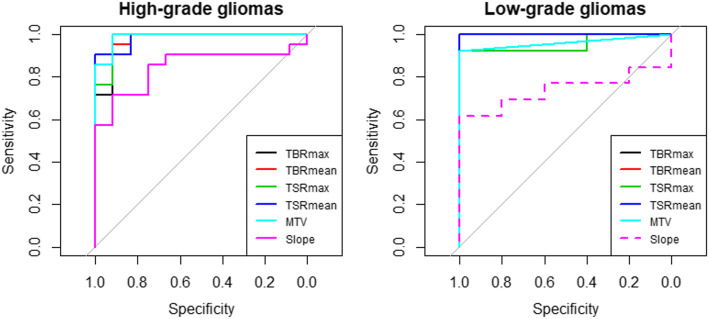
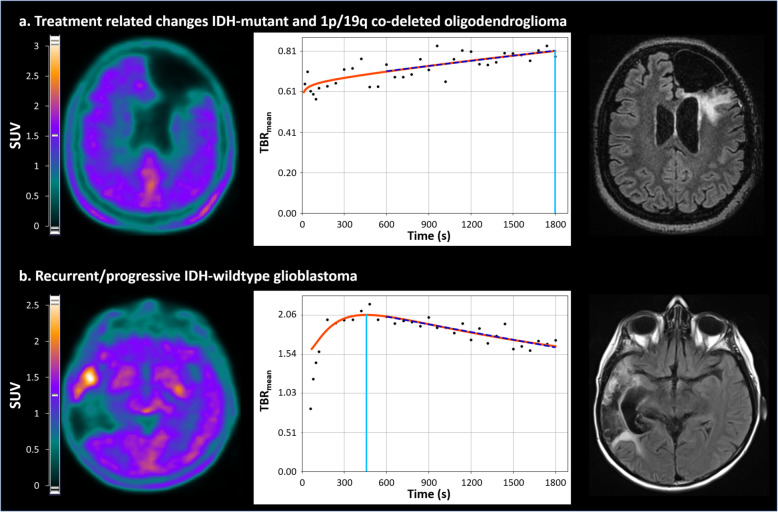
Methods: Fifty-one consecutive patients who underwent an [18F]-F-DOPA PET for a suspected glioma recurrence/progression at post-resection MRI, were retrospectively included. Static parameters, including mean and maximum tumor-to-normal-brain (TBR) ratios, tumor-to-striatum (TSR) ratios, and metabolic tumor volume (MTV), as well as dynamic parameters with time-to-peak (TTP) values and curve slope, were tested for predicting the following: (1) glioma recurrence/progression at 6 months after the PET exam and (2) survival on longer follow-up.

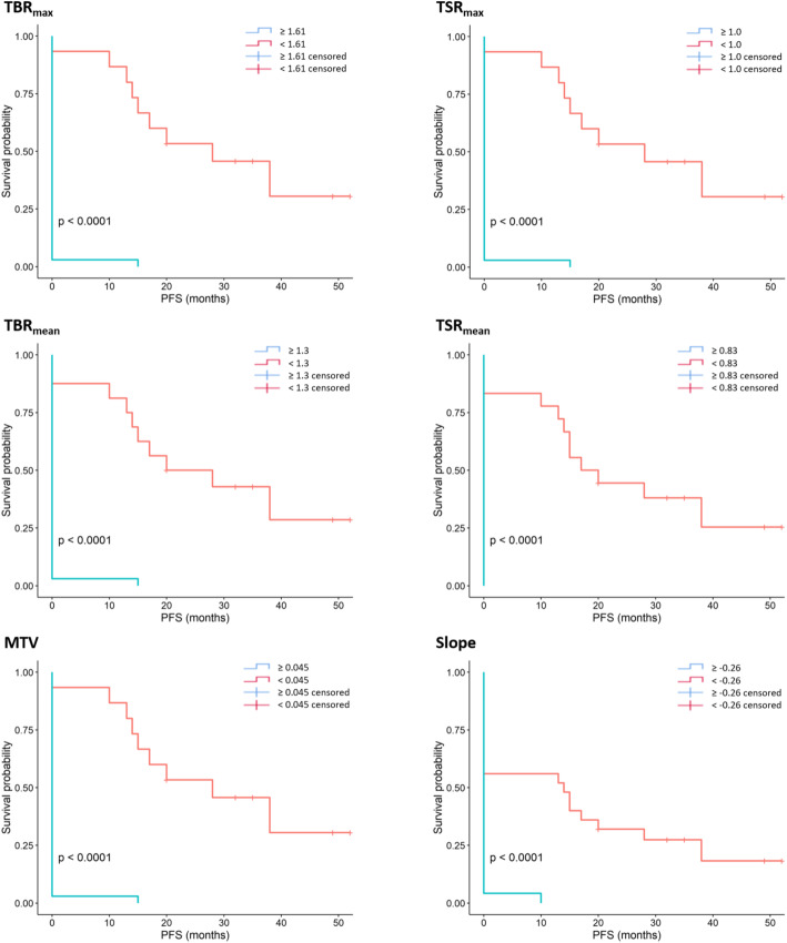
Results: All static parameters were significant predictors of glioma recurrence/progression (accuracy ≥ 94%) with all parameters also associated with mean progression-free survival (PFS) in the overall population (all p < 0.001, 29.7 vs. 0.4 months for TBRmax, TSRmax, and MTV). The curve slope was the sole dynamic PET predictor of glioma recurrence/progression (accuracy = 76.5%) and was also associated with mean PFS (p < 0.001, 18.0 vs. 0.4 months). However, no additional information was provided relative to static parameters in multivariate analysis.

Conclusion: Although patients with glioma recurrence/progression can be detected by both static and dynamic [18F]-F-DOPA PET parameters, most of this diagnostic information can be achieved by conventional static parameters.

Keywords: Amino-acid PET; Dynamic analysis; Glioma; Recurrence; [18F]-F-DOPA.


Conflict of interest statement

All authors declare that they have no competing interests.

Figures

Fig. 1
Fig. 1
Receiver operating characteristic (ROC) curves for TBRmax, TBRmean, TSRmax, TSRmean, MTV, and slope parameters for differentiating between recurrent or progressive gliomas and treatment-related changes in high-grade gliomas (n = 33, left panel) and in low-grade gliomas (n = 18, right panel). Significant ROC curves are represented in solid lines
Fig. 2
Fig. 2
Kaplan-Meier survival plots for the prediction of progression-free survival using maximal tumor-to-background ratios (upper left panel), mean tumor-to-background ratios (middle left panel), metabolic tumor volume (lower left panel), maximal tumor-to-striatum ratio (upper right panel), mean tumor-to-striatum ratio (middle right panel), and slope (lower right panel) as discriminators. Corresponding log-rank test adjusted p values are < 0.0001 for all presented curves
Fig. 3
Fig. 3
Representative examples of patients with or without glioma recurrence/progression investigated with [18F]-F-DOPA PET imaging, with axial slices of [18F]-F-DOPA PET (left column), dynamic TBRmean curves (middle column) providing the time-to-peak delay-time (light blue dotted line), and the 10-to-30 min slope (dark blue dotted line), along with, for illustrative purposes, the same slice location recorded on a FLAIR MRI sequence (right column). a 51-year-old woman with no recurrent or progressive glioma (TBRmean = 0.8, TBRmax = 1.1, TSRmean = 0.5, TSRmax = 0.7, MTV = 0 mL, TTP = 30 min, and slope = 0.31 h−1). b 46-year-old woman with a progressive IDH-wildtype glioblastoma (TBRmean = 1.9, TBRmax = 2.5, TSRmean = 1.2, TSRmax = 1.6, MTV = 6.13 mL, TTP = 7.6 min, and slope = − 1.22 h−1)

References

    1. Ostrom QT, Gittleman H, Fulop J, Liu M, Blanda R, Kromer C, et al. CBTRUS Statistical report: primary brain and central nervous system tumors diagnosed in the United States in 2008-2012. Neuro-Oncol. 2015;17(Suppl 4):iv1–i62. - PMC - PubMed
    1. Lamborn KR, Yung WKA, Chang SM, Wen PY, Cloughesy TF, DeAngelis LM, et al. Progression-free survival: an important end point in evaluating therapy for recurrent high-grade gliomas. Neuro-Oncol. 2008;10:162–170. - PMC - PubMed
    1. Albert NL, Weller M, Suchorska B, Galldiks N, Soffietti R, Kim MM, et al. Response Assessment in Neuro-Oncology working group and European Association for Neuro-Oncology recommendations for the clinical use of PET imaging in gliomas. Neuro-Oncol. 2016;18:1199–1208. - PMC - PubMed
    1. Law I, Albert NL, Arbizu J, Boellaard R, Drzezga A, Galldiks N, et al. Joint EANM/EANO/RANO practice guidelines/SNMMI procedure standards for imaging of gliomas using PET with radiolabelled amino acids and [18F]FDG: version 1.0. Eur J Nucl Med Mol Imaging. 2019;46:540–557. - PMC - PubMed
    1. Chen W, Silverman DHS, Delaloye S, Czernin J, Kamdar N, Pope W, et al. 18F-FDOPA PET imaging of brain tumors: comparison study with 18F-FDG PET and evaluation of diagnostic accuracy. J Nucl Med. 2006;47:904–911. - PubMed

LinkOut - more resources