Review
. 2020 Aug;29(8):1707-1723.
doi: 10.1002/pro.3895. Epub 2020 Jun 26.

These motors were made for walking

Affiliations
Review

These motors were made for walking

Byron Hunter et al. Protein Sci. 2020 Aug.

Abstract

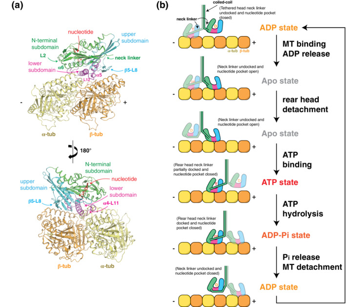
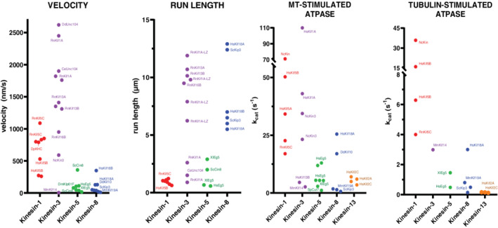
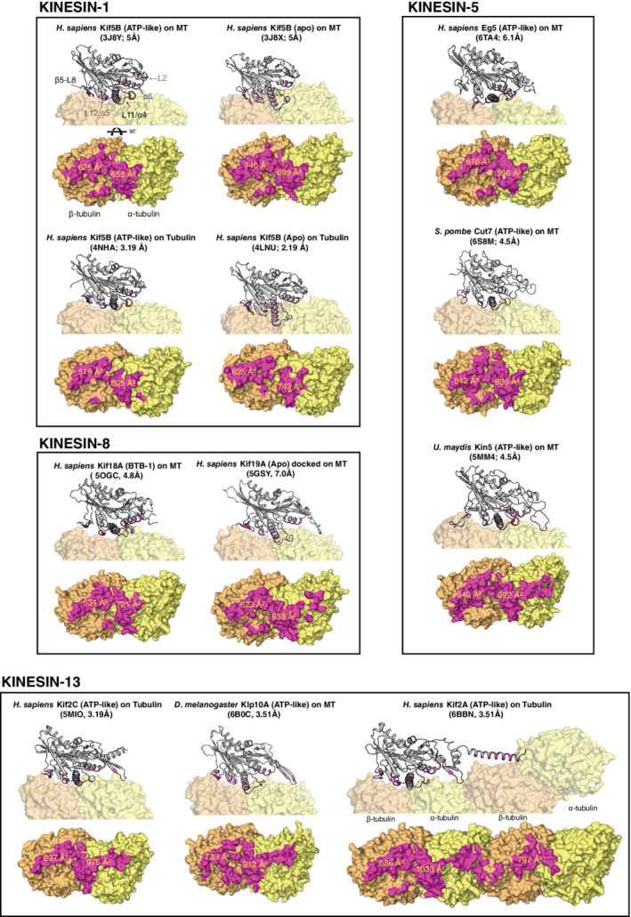
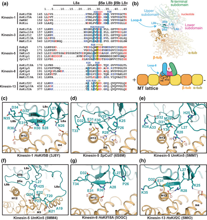
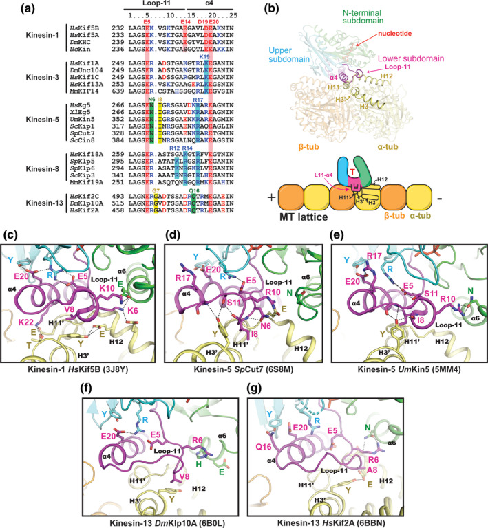
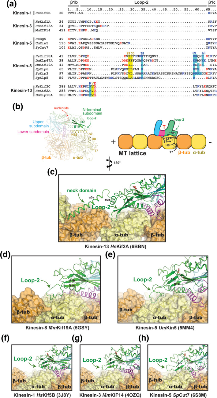
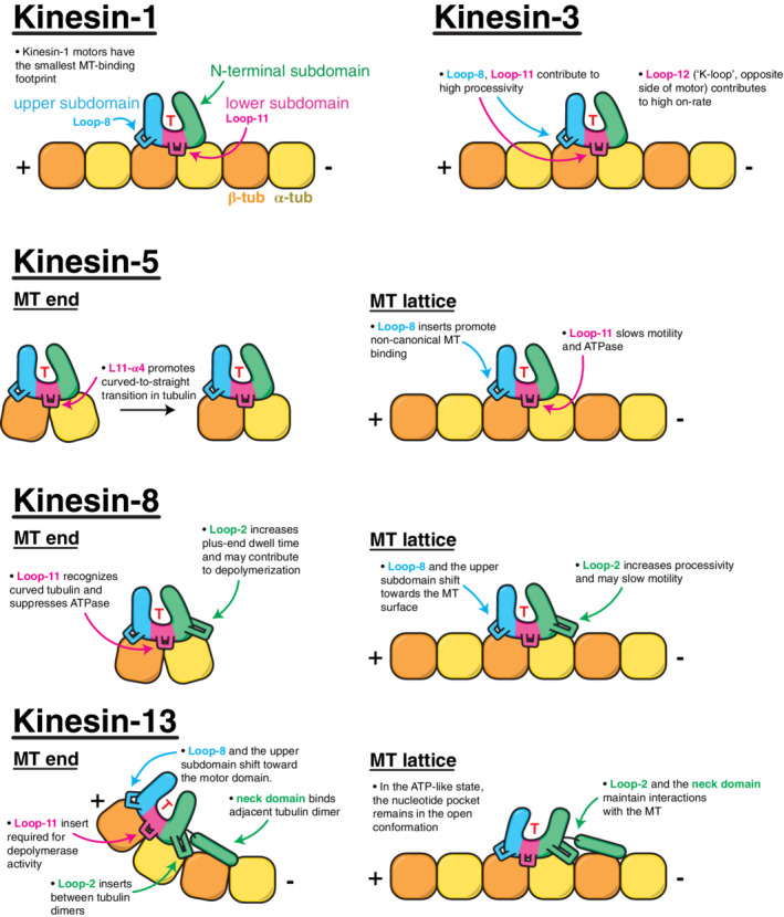
Kinesins are a diverse group of adenosine triphosphate (ATP)-dependent motor proteins that transport cargos along microtubules (MTs) and change the organization of MT networks. Shared among all kinesins is a ~40 kDa motor domain that has evolved an impressive assortment of motility and MT remodeling mechanisms as a result of subtle tweaks and edits within its sequence. Several elegant studies of different kinesin isoforms have exposed the purpose of structural changes in the motor domain as it engages and leaves the MT. However, few studies have compared the sequences and MT contacts of these kinesins systematically. Along with clever strategies to trap kinesin-tubulin complexes for X-ray crystallography, new advancements in cryo-electron microscopy have produced a burst of high-resolution structures that show kinesin-MT interfaces more precisely than ever. This review considers the MT interactions of kinesin subfamilies that exhibit significant differences in speed, processivity, and MT remodeling activity. We show how their sequence variations relate to their tubulin footprint and, in turn, how this explains the molecular activities of previously characterized mutants. As more high-resolution structures become available, this type of assessment will quicken the pace toward establishing each kinesin's design-function relationship.

Keywords: cryo-EM structure; crystal structure; kinesin; microtubules; motor protein; tubulin.


Conflict of interest statement

The authors declare no potential conflict of interest.

Figures

FIGURE 1
FIGURE 1
Kinesin–tubulin interaction and mechanochemical cycle. (a) The kinesin motor domain can be divided into three subdomains that move rigidly during the nucleotide/microtubule binding cycle. Each subdomain forms an interface with tubulin via: (1) loop‐2 and α6 in the N‐terminal subdomain; (2) the loop‐11‐α4‐loop‐12‐α5 (L11‐α4‐L12‐α5) cluster in the lower subdomain; and (3) the β5‐loop‐8 (β5‐L8) lobe in the upper subdomain. (b) A model for the kinesin stepping cycle is depicted for a processive kinesin dimer, whose two heads (motor domains) take turns in stepping toward the MT plus end. First, the tethered, ADP‐bound (D) head is swung around the coiled‐coil junction via docking of the neck‐linker against the rear motor domain. Once the tethered head finds a new tubulin binding site and becomes the lead head, adenosine diphosphate (ADP) is released and this “apo” form of the lead head binds strongly. Adenosine triphosphate (ATP) cannot enter the lead head until the trailing head finishes ATP hydrolysis, release phosphate, and detaches from the MT. Once ATP binding (T) occurs in the lead head, the neck‐linker is able to dock against its motor core, directing the tethered trailing head forward, in the plus end direction. Once this new lead head is firmly bound to tubulin, ATP hydrolysis in the new rear head makes detachment possible
FIGURE 2
FIGURE 2
Motor specifications of kinesin families. Each data point corresponds to a reported value from an independent published experiment. Select motors are labeled. Values were obtained from: kinesin‐1, 33 , 34 , 35 , 36 , 37 , 38 , 39 , 40 , 41 , 42 , 43 , 44 kinesin‐3, 32 , 33 , 34 , 37 , 40 , 42 , 43 , 45 , 46 , 47 , 48 , 49 , 50 , 51 kinesin‐5, 41 , 52 , 53 , 54 , 55 , 56 , 57 , 58 , 59 , 60 , 61 , 62 , 63 , 64 kinesin‐8, 35 , 38 , 65 , 66 , 67 , 68 , 69 , 70 , 71 , 72 , 73 , 74 , 75 , 76 and kinesin‐13. 77 , 78 , 79 Note that several run length values for kinesin‐3 and ‐8 were limited by the length of MTs assembled in vitro
FIGURE 3
FIGURE 3
Microtubule‐binding surfaces for select families of kinesins. Kinesin structures are displayed as cartoons and tubulin is shown as surface representations. Interfaces were identified with ePISA 107 and are colored magenta. Values for buried surface are were calculated by ePISA using coordinate files retrieved from the Protein Data Bank (https://www.rcsb.org/). Nucleotide state, PDB IDs, and the overall map resolution for each structure are shown in parentheses
FIGURE 4
FIGURE 4
Comparison of β5‐L8 lobe structures and MT interactions. (a) Sequence alignment of β5‐L8 segments for selected kinesin family members. Positive and negative‐charged residues are colored blue and red, respectively. (b) Structural and cartoon representation highlighting the location of the β5‐L8 lobe and its interface with tubulin. (c–h) The conformation of the β5‐L8 lobe in each kinesin structure (cyan), and its contact with β‐tubulin (orange), are shown. Residue numbers in the kinesins have been changed to correspond to the columns in the sequence alignment for simplicity. PDB IDs are shown in parentheses. (e, f) Two different conformations of the β5‐L8 lobe that are observed in UmKin5 models (5MM4 and 5MM7)
FIGURE 5
FIGURE 5
Comparison of the L11‐α4 region. (a) Sequence alignment of the L11‐α4 segments for selected kinesin family members. (b) Location of L11‐α4 within the motor domain and its interface with tubulin. (c–g) The conformation of L11‐α4 in each kinesin structure (magenta) and its contacts with α‐tubulin (yellow) are shown. Residue numbers in the kinesins have been changed to correspond to the columns in the sequence alignment for simplicity. PDB IDs are shown in parentheses
FIGURE 6
FIGURE 6
Comparison of loop‐2 structures and MT interactions. (a) Sequence alignment of the loop‐2 region for selected kinesin family members. (b) Location of loop‐2 within the motor domain and its interface with tubulin. (c–h) The conformation of loop‐2 in each kinesin structure (green) and its contacts with tubulin (orange and yellow) are shown. PDB IDs are shown in parentheses. (g) The coordinates for the MmKIF14 motor domain (4OZQ) were docked onto the MT‐bound structure of apo kinesin‐1 (3J8X)
FIGURE 7
FIGURE 7
Summary of kinesin‐family‐specific adaptations and their role in motor activity

References

    1. Saunders WS, Hoyt MA. Kinesin‐related proteins required for structural integrity of the mitotic spindle. Cell. 1992;70:451–458. - PubMed
    1. Roof DM, Meluh PB, Rose MD. Multiple kinesin‐related proteins in yeast mitosis. Cold Spring Harb Symp Quant Biol. 1991;56:693–703. - PubMed
    1. Roof DM, Meluh PB, Rose MD. Kinesin‐related proteins required for assembly of the mitotic spindle. J Cell Biol. 1992;118:95–108. - PMC - PubMed
    1. Woodruff JB, Drubin DG, Barnes G. Mitotic spindle disassembly occurs via distinct subprocesses driven by the anaphase‐promoting complex, aurora B kinase, and kinesin‐8. J Cell Biol. 2010;191:795–808. - PMC - PubMed
    1. Desai A, Mitchison TJ. Microtubule polymerization dynamics. Annu Rev Cell Dev Biol. 1997;13:83–117. - PubMed

Publication types

Grants and funding