. 2020 Aug;35(8):1357-1368.
doi: 10.1002/mds.28063. Epub 2020 May 30.

DNAJC6 Mutations Disrupt Dopamine Homeostasis in Juvenile Parkinsonism-Dystonia

Affiliations

DNAJC6 Mutations Disrupt Dopamine Homeostasis in Juvenile Parkinsonism-Dystonia

Joanne Ng et al. Mov Disord. 2020 Aug.

Abstract

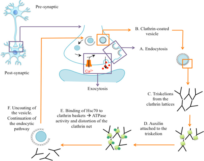
Background: Juvenile forms of parkinsonism are rare conditions with onset of bradykinesia, tremor and rigidity before the age of 21 years. These atypical presentations commonly have a genetic aetiology, highlighting important insights into underlying pathophysiology. Genetic defects may affect key proteins of the endocytic pathway and clathrin-mediated endocytosis (CME), as in DNAJC6-related juvenile parkinsonism.

Objective: To report on a new patient cohort with juvenile-onset DNAJC6 parkinsonism-dystonia and determine the functional consequences on auxilin and dopamine homeostasis.

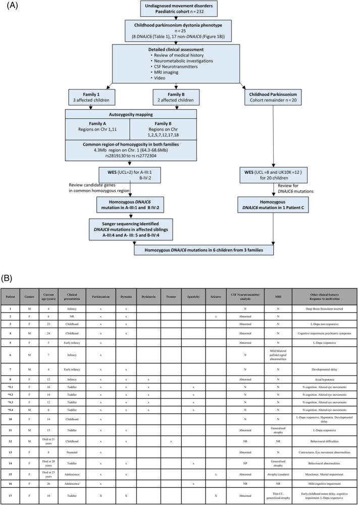
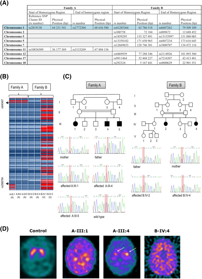
Methods: Twenty-five children with juvenile parkinsonism were identified from a research cohort of patients with undiagnosed pediatric movement disorders. Molecular genetic investigations included autozygosity mapping studies and whole-exome sequencing. Patient fibroblasts and CSF were analyzed for auxilin, cyclin G-associated kinase and synaptic proteins.

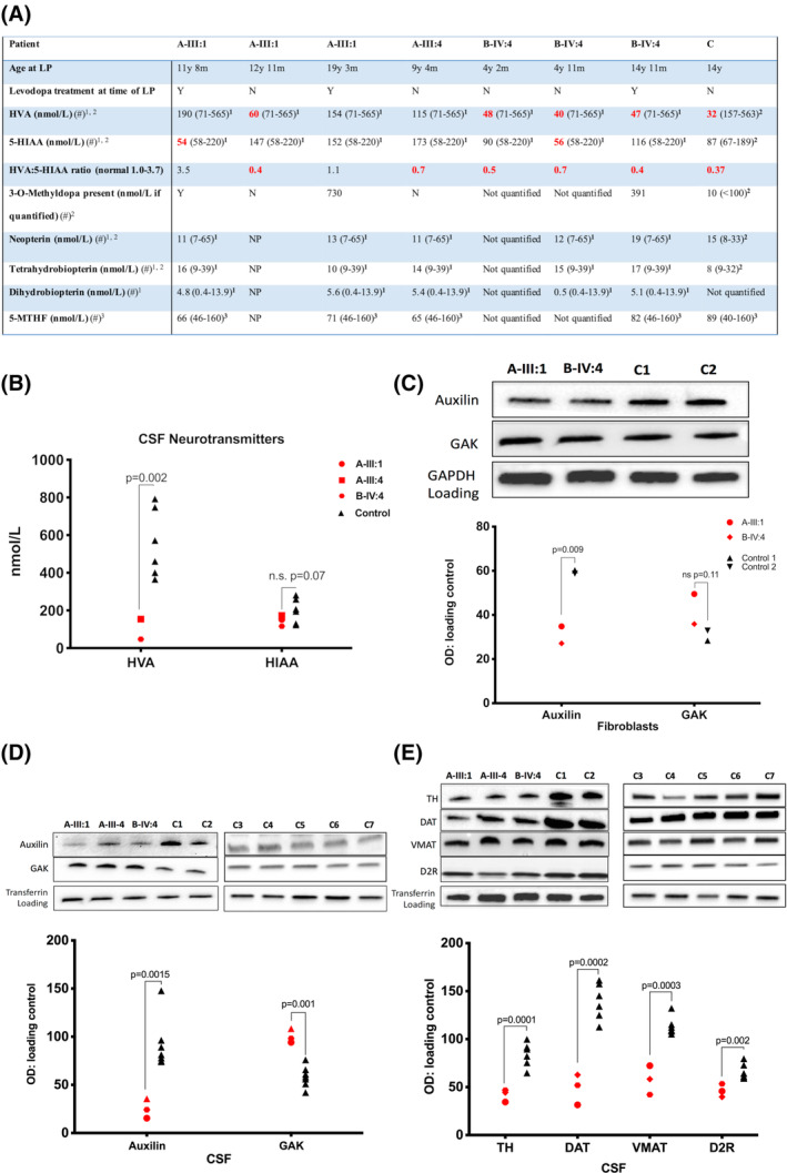
Results: We identified 6 patients harboring previously unreported, homozygous nonsense DNAJC6 mutations. All presented with neurodevelopmental delay in infancy, progressive parkinsonism, and neurological regression in childhood. 123 I-FP-CIT SPECT (DaTScan) was performed in 3 patients and demonstrated reduced or absent tracer uptake in the basal ganglia. CSF neurotransmitter analysis revealed an isolated reduction of homovanillic acid. Auxilin levels were significantly reduced in both patient fibroblasts and CSF. Cyclin G-associated kinase levels in CSF were significantly increased, whereas a number of presynaptic dopaminergic proteins were reduced.

Conclusions: DNAJC6 is an emerging cause of recessive juvenile parkinsonism-dystonia. DNAJC6 encodes the cochaperone protein auxilin, involved in CME of synaptic vesicles. The observed dopamine dyshomeostasis in patients is likely to be multifactorial, secondary to auxilin deficiency and/or neurodegeneration. Increased patient CSF cyclin G-associated kinase, in tandem with reduced auxilin levels, suggests a possible compensatory role of cyclin G-associated kinase, as observed in the auxilin knockout mouse. DNAJC6 parkinsonism-dystonia should be considered as a differential diagnosis for pediatric neurotransmitter disorders associated with low homovanillic acid levels. Future research in elucidating disease pathogenesis will aid the development of better treatments for this pharmacoresistant disorder. © 2020 The Authors. Movement Disorders published by Wiley Periodicals, Inc. on behalf of International Parkinson and Movement Disorder Society.

Keywords: DNAJC6; auxilin; dopamine; dystonia; parkinsonism.


Figures

Figure 1
Figure 1
Juvenile parkinsonism cohort: clinical features and molecular genetic investigation. (A) Flowchart demonstrating the pathway of molecular genetic investigations in a subcohort of 25 children with juvenile parkinsonism. (B) Clinical characteristics of 20 children from 17 families. Early infancy <3 months; infancy 3 to 12 months; toddler 12 to 24 months; childhood 2 to 13 years; adolescence 13 to 18 years. *Consanguineous family. M, male; F, female; N, normal; NP, not performed; NR, not reported. [Color figure can be viewed at wileyonlinelibrary.com]
Figure 2
Figure 2
Molecular genetic investigations and DaTSCAN imaging. (A) Family A and B SNP array results showing homozygous regions detected. For each chromosome, the start and end point is specified using the Reference SNP Cluster ID (rs number) and physical position. (B) Homozygous SNPs are represented in light blue (AA) and dark blue (BB), heterozygous SNPs in red (AB), and “no calls” in white. (C) Sanger Sequencing confirms a homozygous DNAJC6 mutation, c.766C > T (p.R256*), in all affected children of Family A (A‐III:1, A‐III:4, and A‐III:5) and Family B (B‐IV:2, B‐IV:4). Parents are heterozygous carriers. (D) I‐123‐DaTSCAN™ with SPECT imaging in a control subject, Patient A‐III:1 (19 years 3 months), Patient A‐III:4 (11 years 4 months), and Patient B‐IV:4 (17 years). In Patients A‐III:1 and B‐IV:4, DaTSCAN findings indicate virtually complete absence of tracer uptake in the basal ganglia, with very high background activity, suggesting loss of presynaptic dopaminergic terminals, whereas Patient A‐III:4 showed significantly reduced, albeit still visible, uptake in the head of caudate (left better than right, white arrows). [Color figure can be viewed at wileyonlinelibrary.com]
Figure 3
Figure 3
CSF neurotransmitter analysis and patient fibroblast and CSF immunoblotting. (A) CSF neurotransmitter analysis. Age‐related reference ranges indicated in brackets after each value. Red: abnormal result. Gray: borderline result. Symbol (“#”) indicates reference range: 1Keith Hyland, Robert A.H. Surtees, et al. Pediatr Res 1993;34:10–14; 2Keith Hyland, Future Neurol 2006;1:593–603; 3Surtees R, Hyland K. Biochem Med Metab Biol 1990;44:192–199. (B) Scatterplot of CSF HVA and 5‐HIAA levels (nmol/L) measured by high performance liquid chromatography (patient = red shapes, control = black triangles). Medication at time of CSF sampling: A‐III‐1: co‐careldopa, melatonin, glycopyronium; A‐III‐4: none; B‐IV‐4: l‐dopa, pyridoxine; Control 1: none; Control 2: none. Immunoblot of auxilin and GAK in patient fibroblasts (C) and CSF (D) compared to controls. (E) Immunoblot of patient CSF for TH, DAT, VMAT, and D2R protein levels measured compared to controls. Graphs show mean protein percent optical density (OD) normalized to loading control in patients (red) and controls (black). LP, lumbar puncture; y, years; m, months; 5‐MTHF, 5‐methyltetrahydrofolate; NP, not performed. [Color figure can be viewed at wileyonlinelibrary.com]
Figure 4
Figure 4
Schematic representation of DNAJC6‐encoded auxilin protein function. The role of auxilin in synaptic vesicle recycling and endocytic pathway. (A) A nascent clathrin‐coated pit is formed at the presynaptic membrane, followed by membrane invagination; (B) the pit develops into a clathrin‐coated vesicle, where the clathrin lattice consists of (C) clathrin triskelions formed by three crossed ankle regions. (D) Auxilin (pale green square) binds to an exposed domain in the heavy chain of the clathrin. (E) Auxilin facilitates a conformational change in clathrin that allows binding of Hsc70 and by ATPase‐mediated activity, and the clathrin lattice is disrupted and distorted, leading to (F) clathrin disassembly allowing subsequent delivery of cargo neurotransmitters to the membrane or other vesicle in the endocytic pathway. Hsc70, heat shock cognate protein 70. [Color figure can be viewed at wileyonlinelibrary.com]

References

    1. Kitada T, Asakawa S, Hattori N, et al. Mutations in the parkin gene cause autosomal recessive juvenile parkinsonism. Nature 1998;392:605–608. - PubMed
    1. Valente EM, Abou‐Sleiman PM, Caputo V, et al. Hereditary early‐onset Parkinson's disease caused by mutations in PINK1. Science 2004;304:1158–1160. - PubMed
    1. Bonifati V, Rizzu P, van Baren MJ, et al. Mutations in the DJ‐1 gene associated with autosomal recessive early‐onset parkinsonism. Science 2003;299:256–259. - PubMed
    1. Ramirez A, Heimbach A, Gründemann J, et al. Hereditary parkinsonism with dementia is caused by mutations in ATP13A2, encoding a lysosomal type 5 P‐type ATPase. Nat Genet 2006;38:1184–1191. - PubMed
    1. Paisan‐Ruiz C, Bhatia KP, Li A, et al. Characterization of PLA2G6 as a locus for dystonia‐parkinsonism. Ann Neurol 2009;65:19–23. - PMC - PubMed

Publication types