Review
. 2020 Sep:163:103617.
doi: 10.1016/j.mod.2020.103617. Epub 2020 May 27.

Mouse gastrulation: Coordination of tissue patterning, specification and diversification of cell fate

Affiliations
Review

Mouse gastrulation: Coordination of tissue patterning, specification and diversification of cell fate

Evan S Bardot et al. Mech Dev. 2020 Sep.

Abstract

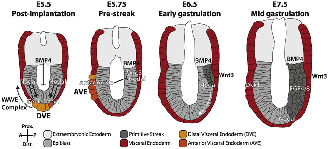
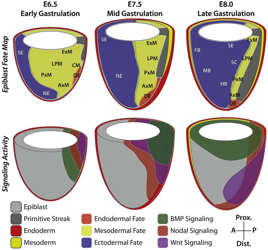
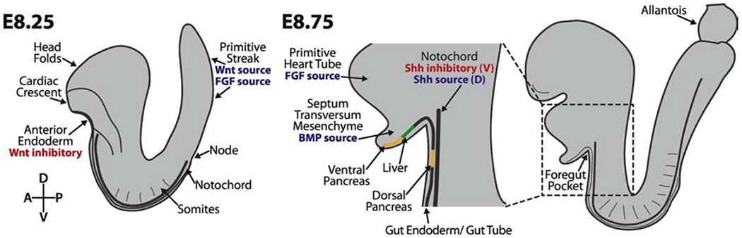
During mouse embryonic development a mass of pluripotent epiblast tissue is transformed during gastrulation to generate the three definitive germ layers: endoderm, mesoderm, and ectoderm. During gastrulation, a spatiotemporally controlled sequence of events results in the generation of organ progenitors and positions them in a stereotypical fashion throughout the embryo. Key to the correct specification and differentiation of these cell fates is the establishment of an axial coordinate system along with the integration of multiple signals by individual epiblast cells to produce distinct outcomes. These signaling domains evolve as the anterior-posterior axis is established and the embryo grows in size. Gastrulation is initiated at the posteriorly positioned primitive streak, from which nascent mesoderm and endoderm progenitors ingress and begin to diversify. Advances in technology have facilitated the elaboration of landmark findings that originally described the epiblast fate map and signaling pathways required to execute those fates. Here we will discuss the current state of the field and reflect on how our understanding has shifted in recent years.

Keywords: Axis patterning; Cell fate specification; Gastrulation; Mouse development; Organogenesis.


Figures

Figure 1:
Figure 1:. Anterior-posterior axis patterning positions the primitive streak prior to gastrulation.
After implantation, the embryo transitions from using the proximal-distal axis to the embryonic anterior-posterior (AP) axis. The establishment of this axis depends on a sequence of events including specification of the DVE at E5.5 and formation and migration (dashed black arrow) of the AVE at E5.75. The primitive streak is restricted to the posterior pole by E6.5 and elongates distally and anteriorly (white dashed arrow) until E7.5. Solid arrows indicate activation, bar-headed lines indicate inhibition. Adapted from (Rivera-Pérez and Hadjantonakis, 2015).
Figure 2:
Figure 2:. Dynamic signaling in the gastrulating mouse embryo influences epiblast cell fate decisions.
Regions of the epiblast have been shown to have reproducible lineage destinies, resulting in fate maps for multiple stages during gastrulation (top row). These fates are determined by the signaling environment cells are exposed to as they ingress through and migrate away from the primitive streak (bottom row). Thus, the combination of fate and signaling maps are instructive for how specific lineages are specified during development. DE, definitive endoderm; CM, cardiac mesoderm; ExM, extraembryonic mesoderm; LPM, lateral plate mesoderm; PxM, paraxial mesoderm, AxM, axial mesoderm; SE, surface ectoderm; SC, spinal cord; NE, neurectoderm; FB, forebrain; MB, midbrain; HB, hindbrain.
Figure 3:
Figure 3:. Differentiation of early organ progenitors occurs in an interdependent manner.
Cells fated to give rise to individual organs arrive at their destinations at different times, and are therefore exposed to different signaling environments. Late specified cells and axial progenitors are exposed to Wnt and FGF signals at the posterior of the embryo. Heart progenitors form the cardiac crescent at E8.25 adjacent to the Wnt-inhibitory anterior endoderm. As these cells differentiate and form the primitive heart tube, they express FGF ligands. These signals, in combination with BMP from the septum transversum mesenchyme, act on the foregut endoderm to induce liver fates and inhibit pancreas formation. Foregut endoderm just ventral to that domain will become the ventral pancreas. The notochord lies between the gut tube endoderm and the neural tube. The notochord secretes FGF and TGF-β to inhibit Shh expression in the gut tube endoderm, which is necessary for the formation of the dorsal pancreas, but is an important Shh source for specifying cell fates within the ventral neural tube.

References

    1. Abe T, Kutsuna N, Kiyonari H, Furuta Y, Fujimori T, 2018. ROSA26 reporter mouse lines and image analyses reveal distinct region-specific cell behaviors in the visceral endoderm. Dev. 145 10.1242/dev.165852 - DOI - PubMed
    1. Acloque H., Ocaña OH, Matheu A, Rizzoti K, Wise C, Lovell-Badge R, Nieto MA, 2011. Reciprocal repression between Sox3 and Snail transcription factors defines embryonic territories at gastrulation. Dev. Cell. 10.1016/j.devcel.2011.07.005 - DOI - PMC - PubMed
    1. Alemany A, Florescu M, Baron CS, Peterson-Maduro J, Van Oudenaarden A, 2018. Whole-organism clone tracing using single-cell sequencing. Nature 556, 108–112. 10.1038/nature25969 - DOI - PubMed
    1. Ang SL, Rossant J, 1994. HNF-3β is essential for node and notochord formation in mouse development. Cell 78, 561–574. 10.1016/0092-8674(94)90522-3 - DOI - PubMed
    1. Antonica F, Orietti LC, Mort RL, Zernicka-Goetz M, 2019. Concerted cell divisions in embryonic visceral endoderm guide anterior visceral endoderm migration. Dev. Biol 450, 132–140. 10.1016/j.ydbio.2019.03.016 - DOI - PMC - PubMed

Publication types

LinkOut - more resources