

Clonal lineage from normal endometrium to ovarian clear cell carcinoma through ovarian endometriosis
- PMID: 32473611
- PMCID: PMC7419022
- DOI: 10.1111/cas.14507
Clonal lineage from normal endometrium to ovarian clear cell carcinoma through ovarian endometriosis
Abstract
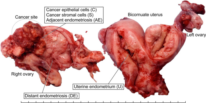
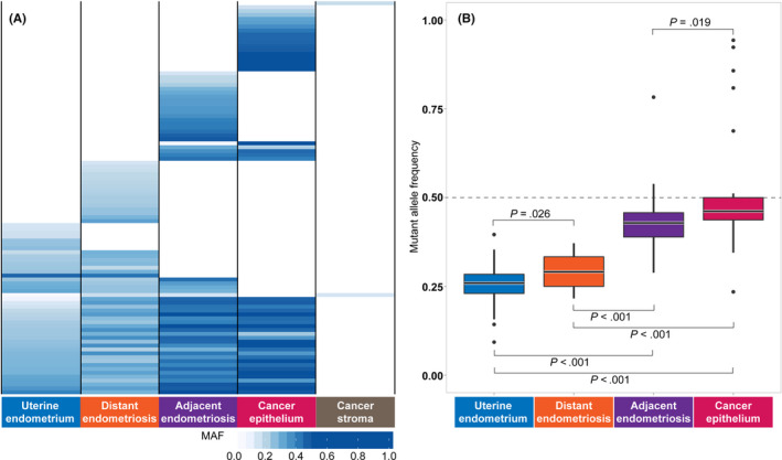
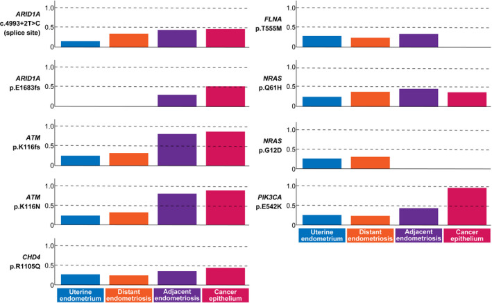
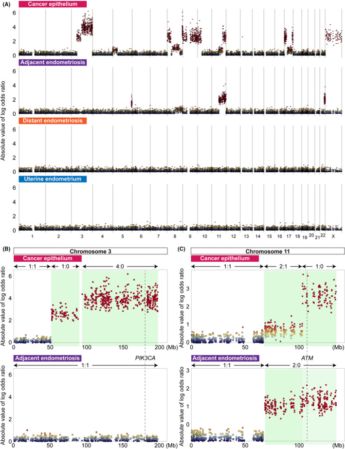
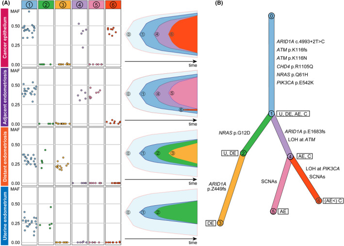
Clear cell carcinoma of the ovary is thought to arise from endometriosis. In addition, retrograde menstruation of shed endometrium is considered the origin of endometriosis. However, little evidence supports cellular continuity from uterine endometrium to clear cell carcinoma through endometriosis at the genomic level. Here, we performed multiregional whole-exome sequencing to clarify clonal relationships among uterine endometrium, ovarian endometriosis and ovarian clear cell carcinoma in a 56-year-old patient. Many somatic mutations including cancer-associated gene mutations in ARID1A, ATM, CDH4, NRAS and PIK3CA were shared among epithelium samples from uterine endometrium, endometriotic lesions distant from and adjacent to the carcinoma, and the carcinoma. The mutant allele frequencies of shared mutations increased from uterine endometrium to distant endometriosis, adjacent endometriosis, and carcinoma. Although a splice site mutation of ARID1A was shared among the four epithelium samples, a frameshift insertion in ARID1A was shared by adjacent endometriosis and carcinoma samples, suggesting that the biallelic mutations triggered malignant transformation. Somatic copy number alterations, including loss of heterozygosity events at PIK3CA and ATM, were identified only in adjacent endometriosis and carcinoma, suggesting that mutant allele-specific imbalance is another key factor driving malignant transformation. By reconstructing a clonal evolution tree based on the somatic mutations, we showed that the epithelium samples were derived from a single ancestral clone. Although the study was limited to a single patient, the results from this illustrative case could suggest the possibility that epithelial cells of ovarian endometriosis and clear cell carcinoma were descendants of uterine endometrial epithelium.
Keywords: clonal evolution; endometriosis; endometrium; genome; ovarian neoplasms.
© 2020 The Authors. Cancer Science published by John Wiley & Sons Australia, Ltd on behalf of Japanese Cancer Association.
Conflict of interest statement
The authors declare that they have no conflicts of interest.
Figures
Similar articles
-
Frequent PIK3CA mutations in eutopic endometrium of patients with ovarian clear cell carcinoma.Mod Pathol. 2021 Nov;34(11):2071-2079. doi: 10.1038/s41379-021-00861-3. Epub 2021 Jun 25. Mod Pathol. 2021. PMID: 34172890 Free PMC article.
-
Different mutation profiles between epithelium and stroma in endometriosis and normal endometrium.Hum Reprod. 2019 Oct 2;34(10):1899-1905. doi: 10.1093/humrep/dez155. Hum Reprod. 2019. PMID: 31621846
-
Clonal Expansion and Diversification of Cancer-Associated Mutations in Endometriosis and Normal Endometrium.Cell Rep. 2018 Aug 14;24(7):1777-1789. doi: 10.1016/j.celrep.2018.07.037. Cell Rep. 2018. PMID: 30110635
-
Endometriosis-associated Ovarian Cancers.Clin Obstet Gynecol. 2017 Dec;60(4):711-727. doi: 10.1097/GRF.0000000000000320. Clin Obstet Gynecol. 2017. PMID: 28990985 Review.
-
Malignant transformation of endometriosis: application of laser microdissection for analysis of genetic alterations according to pathological changes.Med Electron Microsc. 2004 Jun;37(2):97-100. doi: 10.1007/s00795-003-0233-0. Med Electron Microsc. 2004. PMID: 15221651 Review.
Cited by
-
Spatiotemporal dynamics of clonal selection and diversification in normal endometrial epithelium.Nat Commun. 2022 Feb 17;13(1):943. doi: 10.1038/s41467-022-28568-2. Nat Commun. 2022. PMID: 35177608 Free PMC article.
-
Establishment of a Novel In Vitro Model of Endometriosis with Oncogenic KRAS and PIK3CA Mutations for Understanding the Underlying Biology and Molecular Pathogenesis.Cancers (Basel). 2021 Jun 25;13(13):3174. doi: 10.3390/cancers13133174. Cancers (Basel). 2021. PMID: 34202354 Free PMC article.
-
How Does Endometriosis Lead to Ovarian Cancer? The Molecular Mechanism of Endometriosis-Associated Ovarian Cancer Development.Cancers (Basel). 2021 Mar 22;13(6):1439. doi: 10.3390/cancers13061439. Cancers (Basel). 2021. PMID: 33809880 Free PMC article. Review.
-
Frequent PIK3CA mutations in eutopic endometrium of patients with ovarian clear cell carcinoma.Mod Pathol. 2021 Nov;34(11):2071-2079. doi: 10.1038/s41379-021-00861-3. Epub 2021 Jun 25. Mod Pathol. 2021. PMID: 34172890 Free PMC article.
-
Whole-Exome Sequencing of Rare Site Endometriosis-Associated Cancer.Diseases. 2021 Feb 4;9(1):14. doi: 10.3390/diseases9010014. Diseases. 2021. PMID: 33557369 Free PMC article.
References
-
- Suda K, Nakaoka H, Yoshihara K, et al. Clonal expansion and diversification of cancer‐associated mutations in endometriosis and normal endometrium. Cell Rep. 2018;24:1777‐1789. - PubMed
MeSH terms
Grants and funding
LinkOut - more resources
Full Text Sources
Medical
Research Materials
Miscellaneous
