. 2020 May 30;19(1):98.
doi: 10.1186/s12943-020-01217-2.

LINC00173.v1 promotes angiogenesis and progression of lung squamous cell carcinoma by sponging miR-511-5p to regulate VEGFA expression

Affiliations

LINC00173.v1 promotes angiogenesis and progression of lung squamous cell carcinoma by sponging miR-511-5p to regulate VEGFA expression

Jiarong Chen et al. Mol Cancer. .

Abstract

Background: Anti-angiogenic therapy represents a promising strategy for non-small-cell lung cancer (NSCLC) but its application in lung squamous cell carcinoma (SQC) is limited due to the high-risk adverse effects. Accumulating evidence indicates that long noncoding RNAs (lncRNAs) mediate in tumor progression by participating in the regulation of VEGF in NSCLC, which might guide the development of new antiangiogenic strategies.

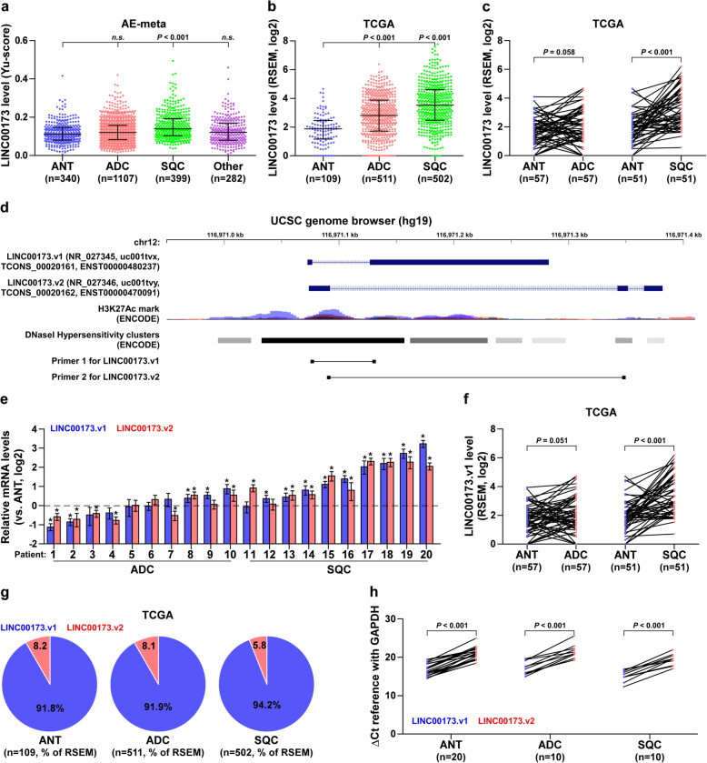
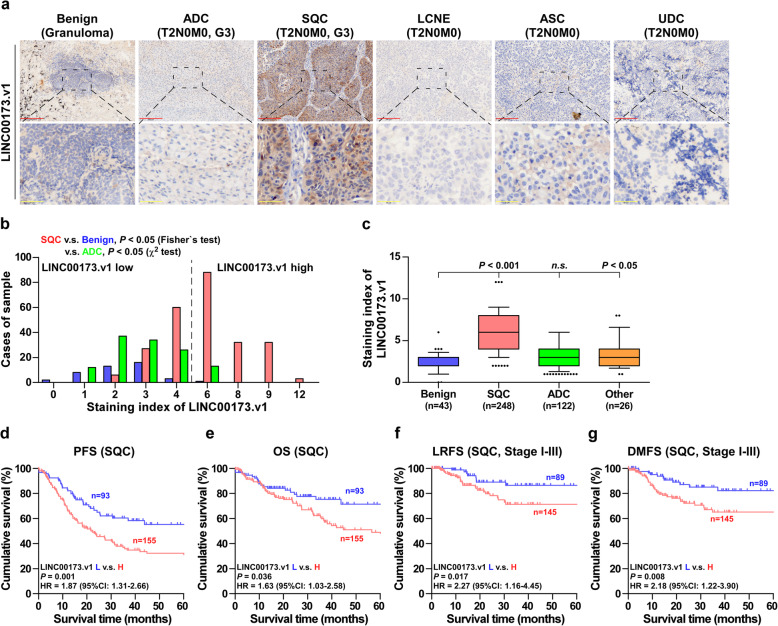
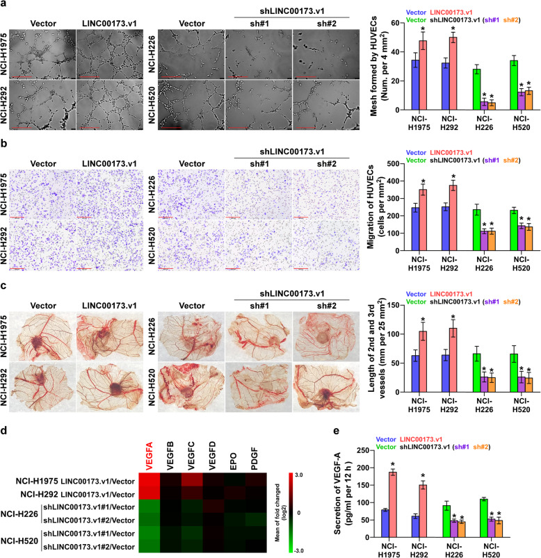
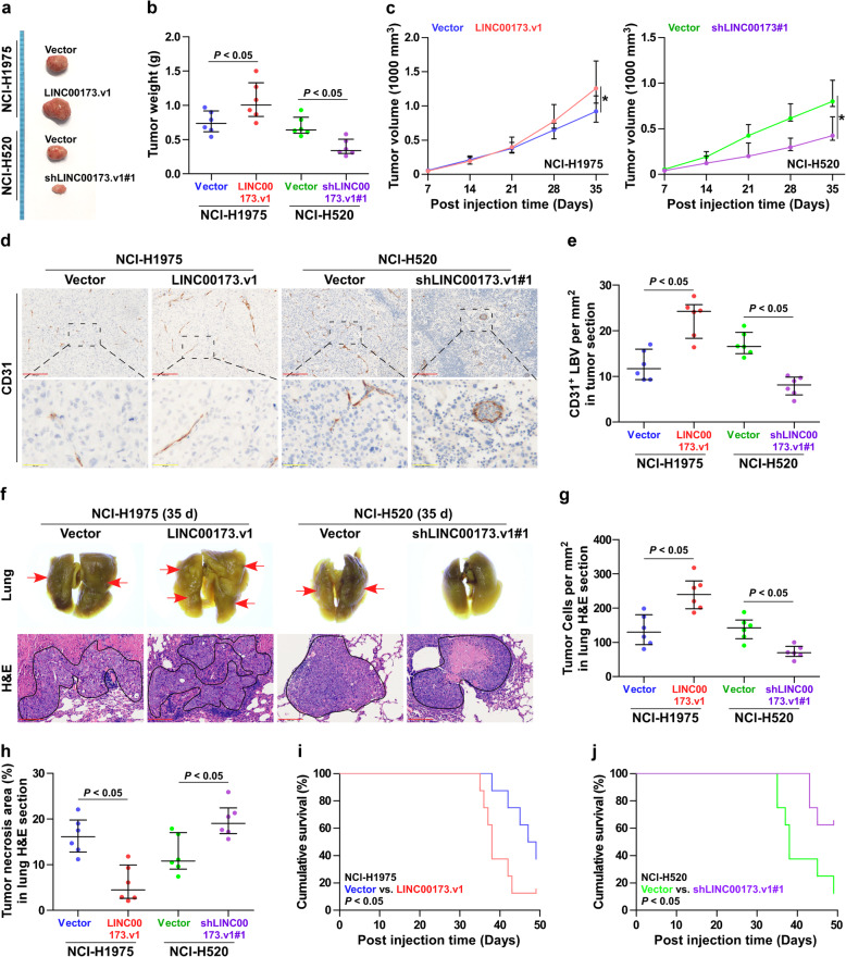
Methods: Differential lncRNA expression in SQC was analyzed in AE-meta and TCGA datasets, and further confirmed in lung cancer tissues and adjacent normal tissues with RT-qPCR and in-situ hybridization. Statistical analysis was performed to evaluate the clinical correlation between LINC00173.v1 expression and survival characteristics. A tube formation assay, chick embryo chorioallantoic membrane assay and animal experiments were conducted to detect the effect of LINC00173.v1 on the proliferation and migration of vascular endothelial cells and tumorigenesis of SQC in vivo. Bioinformatics analysis, RNA immunoprecipitation and luciferase reporter assays were performed to elucidate the downstream target of LINC00173.v1. The therapeutic efficacy of antisense oligonucleotide (ASO) against LINC00173.v1 was further investigated in vivo. Chromatin immunoprecipitation and high throughput data processing and visualization were performed to identify the cause of LINC00173.v1 overexpression in SQC.

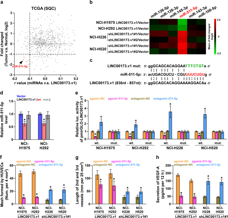
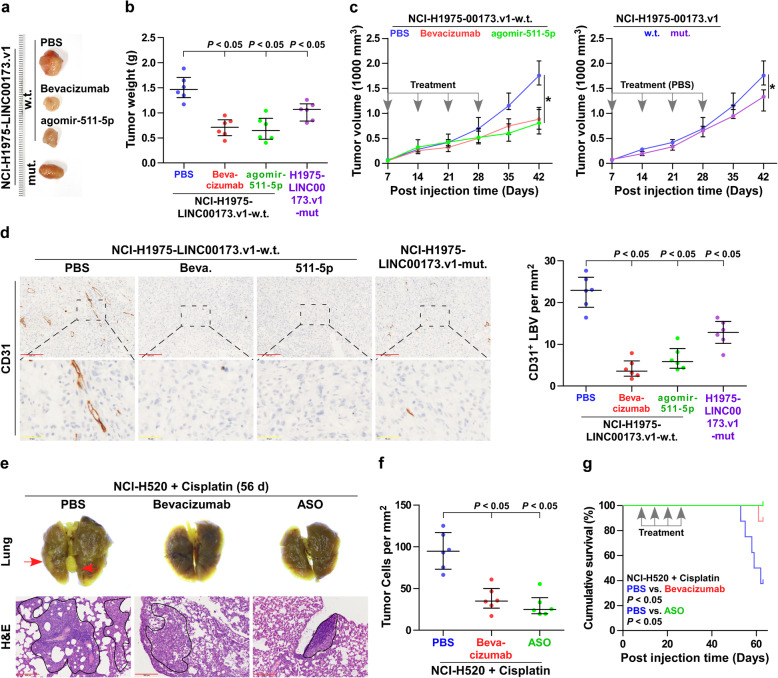
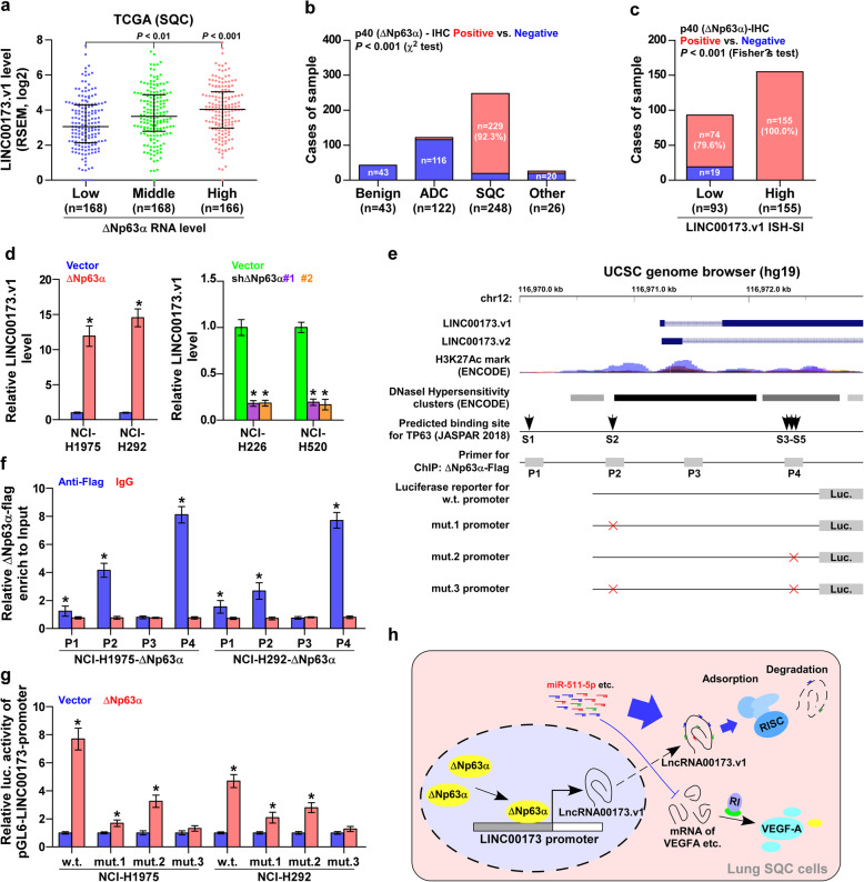
Results: LINC00173.v1 was specifically upregulated in SQC tissues, which predicted poorer overall and progression-free survival in SQC patients. Overexpression of LINC00173.v1 promoted, while silencing LINC00173.v1 inhibited the proliferation and migration of vascular endothelial cells and the tumorigenesis of SQC cells in vitro and in vivo. Our results further revealed that LINC00173.v1 promoted the proliferation and migration of vascular endothelial cells and the tumorigenesis of SQC cells by upregulating VEGFA expression by sponging miR-511-5p. Importantly, inhibition of LINC00173.v1 via the ASO strategy reduced the tumor growth of SQC cells, and enhanced the therapeutic sensitivity of SQC cells to cisplatin in vivo. Moreover, our results showed that squamous cell carcinoma-specific factor ΔNp63α contributed to LINC00173.v1 overexpression in SQC.

Conclusion: Our findings clarify the underlying mechanism by which LINC00173.v1 promotes the proliferation and migration of vascular endothelial cells and the tumorigenesis of SQC, demonstrating that LINC00173.v1-targeted drug in combination with cisplatin may serve as a rational regimen against SQC.

Keywords: Angiogenesis; LINC00173.v1; Lung squamous cell carcinoma; VEGFA; miR-511-5p.


Conflict of interest statement

No conflicts of interest were declared.

Figures

Fig. 1
Fig. 1
LINC00173.v1 is specifically overexpressed in lung squamous cell carcinoma. a Expression level of LINC00173 in 340 adjacent normal tissues (ANT), 1107 adenocarcinoma cell carcinoma (ADC), 399 squamous cell carcinoma (SQC) and 282 other subtypes of lung cancer, including Adenosquamous carcinoma (ASC), Large cell neuroendocrine carcinoma (LCNE), Undifferentiated carcinoma (UDC), Epidermoid carcinoma (EPC), Sarcomatoid carcinoma (SARC), Pleomorphic carcinoma (PMC), and Non-specific types of NSCLC in AE-meta. Each bar represents the median values ± quartile values. P value was determined by one-way ANOVA test. n.s. indicates no significance. b Expression level of LINC00173 in 109 ANT, 511 ADC and 502 SQC in TCGA. Each bar represents the median values ± quartile values. P value was determined by one-way ANOVA test. c Comparison of LINC00173 expression between SQC, ADC and their matched ANT in TCGA. *P < 0.05 by paired t test. d Identification of two LINC00173 transcripts and their exon sequences in UCSC genome browser. e RNA expression of LINC00173.v1 and LINC00173.v2 in ADC and SQC patient samples. *P < 0.05 by paired t test. f Comparison of LINC00173.v1 expression between ADC, SQC and matched ANT in TCGA. *P < 0.05 by paired t test. g RSEM percentages of LINC00173.v1 and LINC00173.v2 based on their reads in lung cancer dataset from TCGA. h ΔCt references of LINC00173.v1 and LINC00173.v2 in lung cancer tissues and ANT in real-time PCR analysis relative to GAPDH. *P < 0.05 by paired t test
Fig. 2
Fig. 2
Overexpression of LINC00173.v1 predicts early relapse and metastasis in SQC. a Representative sections of LINC00173.v1 in 43 benign lung tissues (eg. granuloma), 248 SQC tissues, 122 ADC tissues and 26 other subtypes of lung cancer including LCNE, ASC, and UDC, using in situ hybridization. Scale bars of 100× magnification, 200 μm and 400× magnification, 50 μm. b The number of 43 benign lung tissues, 248 SQC tissues and 122 ADC tissues stratified by staining index of LINC00173.v1. P value was determined by Fisher’s test or χ2 test. c Staining index of LINC00173.v1 in 43 benign lung granuloma, 248 SQC tissues, 122 ADC tissues and 26 other subtypes of lung cancer tissues. Each bar represents the median values ± quartile values. P value was determined by one-way ANOVA test. n.s. indicates no significance. d to g Kaplan–Meier analysis of progression-free survivals (PFS) (d), overall survival (OS) (e), local relapse-free survival (LRFS) (f) and distant metastasis-free survival (DMFS) (g) in SQC patients with low LINC00173.v1 expression versus high LINC00173.v1 expression. P value was determined by Log-rank test. HR indicates hazard ratio; 95%CI indicates 95% confidence interval
Fig. 3
Fig. 3
Silencing LINC00173.v1 represses proliferation and migration of vascular endothelial cells. a Effect of LINC00173.v1 expression in lung cancer cells on human umbilical vein endothelial cells (HUVECs) in tube formation assay. Each bar represents the mean values ± SD of three independent experiments. *P < 0.05 by unpaired t test or one-way ANOVA test. Scale bars, 200 μm. b Effect of LINC00173.v1 expression in lung cancer cells on migration ability of HUVECs in transwell assay. Each bar represents the mean values ± SD of three independent experiments. *P < 0.05 by unpaired t test or one-way ANOVA test. Scale bars, 200 μm. c Effect of LINC00173.v1 on length of the 2nd and 3rd vessels on chick embryo chorioallantoic membrane (CAM). Each bar represents the mean values ± SD of three independent experiments. *P < 0.05 by unpaired t test or one-way ANOVA test. d Real-time PCR analysis of the effect of LINC00173.v1 on expression of multiple proliferation and migration of vascular endothelial cells-associated (angiogenesis-associated) genes. Pseudo-color scale values were log2 transformed. Transcript levels were normalized by GAPDH expression. The experiment was independently performed three times. e Effect of LINC00173.v1 on secretion level of VEGF-A in lung cancer cells by enzyme linked immunosorbent assay (ELISA). Each bar represents the mean values ± SD of three independent experiments. *P < 0.05 by unpaired t test or one-way ANOVA test
Fig. 4
Fig. 4
Silencing LINC00173.v1 inhibits tumorigenesis in lung cancer cells. a Images of the representative excised tumors from the mice at 35 days after injection with the indicated cells. b Average weight of excised tumors from the indicated mice (n = 6). Each bar represents the median values ± quartile values. P value was determined by unpaired t test. c Tumor volumes (n = 6) were measured every 7 days. Each bar represents the median values ± quartile values. *P < 0.05 by unpaired t test in final measurement. d and e Representative sections and CD31+ lymphatic or blood vessel (LBV) density in tumor tissues from the indicated mice groups (n = 6). after 5 weeks of cell injection. Each bar represents the median values ± quartile values. P value was determined by unpaired t test. Scale bars of 100× magnification, 200 μm and 400× magnification, 50 μm. f Metastatic lung tumor nests (red arrow) in the indicated mice group. Lung metastatic tumor tissues in mice were confirmed by H&E staining. Scale bars, 200 μm. g and h Tumor cell number per mm2 and tumor necrotic area in lung H&E section from the indicated mice groups after 5 weeks of tail veins injection. Each bar represents the median values ± quartile values. P value was determined by unpaired t test. i and j Kaplan–Meier analysis of the effect of LINC00173.v1 overexpression (i) and downexpression (j) on cumulative survival in the indicated mice groups (n = 8). P value was determined by Log-rank test
Fig. 5
Fig. 5
LINC00173.v1 functions as a competitive endogenous RNA for miR-511-5p. a Volcano plot analyzed the clinical correlation of LINC00173.v1 with all reported miRNAs in SQC dataset from TCGA (n = 502). The red colors represent significantly and negatively correlated miRNAs with fold change < 0.5 and r value < − 0.1. b Real-time PCR analysis of the effect of LINC00173.v1 on multiple miRNAs expression in the indicated cells. Pseudo-color scale values were log2 transformed. Transcript levels were normalized by U6 expression. The experiment was independently performed three times. c Potential binding sites of mutant and wild-type LINC00173.v1 on miR-511-5p. d Real-time PCR analysis of miR-511-5p expression compared with mutant and wild-type LINC00173.v1. Transcript levels were normalized by U6 expression. Each bar represents the mean values ± SD of three independent experiments. *P < 0.05 by one-way ANOVA test. e Luciferase reporter activity of the influence of miR-511-5p on LINC00173.v1. Each bar represents the mean values ± SD of three independent experiments. *P < 0.05 by one-way ANOVA test. f Effect of miR-511-5p on human umbilical vein endothelial cells (HUVECs) in tube formation assay in the indicated lung cancer cells. Each bar represents the mean values ± SD of three independent experiments. *P < 0.05 by unpaired t test. g Effect of miR-511-5p on length of the 2nd and 3rd vessels on chick embryo chorioallantoic membrane (CAM) in the indicated lung cancer cells. Each bar represents the mean values ± SD of three independent experiments. *P < 0.05 by unpaired t test. h Influence of miR-511-5p on secretion level of VEGF-A in the indicated lung cancer cells. Each bar represents the mean values ± SD of three independent experiments. *P < 0.05 by unpaired t test
Fig. 6
Fig. 6
miR-511-5p/VEGFA axis is essential for LINC00173.v1-induced tumorigenesis in vivo. a Images of the representative excised tumors from the indicated mice at 42 days after injection. b Average weight of excised tumors from the indicated mice (n = 6). Each bar represents the median values ± quartile values. P value was determined by one-way ANOVA test. c Tumor volumes were measured every 7 days. Each bar represents the median values ±quartile values. P value was determined by ANOVA test in final measurement. d Representative sections and CD31+ lymphatic or blood vessel (LBV) in tumor tissues from the indicated mice groups (n = 6) with bevacizumab treatment, agomir-511-5p treatment and the mutant LINC00173.v1-overexpressing NCI-H1975 cells (Left panel). Density of CD31+ lymphatic or blood vessel (LBV) in tumor tissues from the indicated mice groups (Right panel). Each bar represents the median values ± quartile values. P value was determined by one-way ANOVA test. Scale bars of 100× magnification, 200 μm and 400× magnification, 50 μm. e Representative lung nodules (red arrow), lung H&E section and tumor cells in lung sections of therapeutic efficacy of bevacizumab and LINC00173.v1 antisense oligonucleotide (ASO) from the indicated mice groups treated with cisplatin. PBS, bevacizumab and LINC00173.v1 ASO were injected intraperitoneally once weekly for consecutive 4 weeks respectively after 1 week of cell inoculation. Scale bars, 200 μm. f Tumor cell number per mm2 in lung H&E section from the indicated mice groups after 8 weeks of tail veins injection. Each bar represents the median values ± quartile values. P value was determined by one-way ANOVA test. g Kaplan–Meier analysis of the effect of PBS, bevacizumab and LINC00173.v1 ASO on cumulative survival in the indicated mice groups (n = 8). P value was determined by Log-rank test
Fig. 7
Fig. 7
Squamous cell carcinoma-specific factor ΔNp63α contributes to LINC00173.v1 overexpression in SQC. a LINC00173.v1 expression levels in lung cancer tissues with low (n = 168), middle (n = 168) and high (n = 166) ΔNp63α expression from TCGA SQC dataset. Each bar represents the median values ± quartile values. P value was determined by one-way ANOVA test. b Positive percentage of ΔNp63α staining in 43 benign lung granuloma, 248 SQC tissues, 122 ADC tissues and 26 other subtypes of NSCLC. P value was determined by χ2 test. c Cases of sample of immunohistochemical staining of ΔNp63α in SQC tissues stratified by low (n = 93) and high (n = 155) LINC00173.v1 expression levels. P value was determined by Fisher’s test. d Real-time PCR analysis of LINC00173.v1 expression in lung cancer cell lines with changed expression levels of ΔNp63α. Each bar represents the mean values ± SD of three independent experiments. *P < 0.05 by unpaired t test or one-way ANOVA test. e ΔNp63α-binding motifs in the putative promoter region of LINC00173.v1 in UCSC genome browser. f ChIP analysis of the binding sites of ΔNp63α in the promoter region of LINC00173.v1 in lung cancer cells. IgG was used as negative control. Each bar represents the mean values ± SD of three independent experiments. *P < 0.05 by unpaired t test. g Luciferase reporter activity of LINC00173.v1 promoter in ΔNp63α-overexpressing lung cancer cells. Each bar represents the mean values ± SD of three independent experiments. *P < 0.05 by unpaired t test. h Hypothetical model illustrating that squamous cell carcinoma-specific factor ΔNp63α contributes to LINC00173.v1 overexpression in SQC tissues, which further promotes proliferation and migration of vascular endothelial cells and progression of lung squamous cell carcinoma through sponging miR-511-5p to regulate VEGFA expression

References

    1. Bray F, Ferlay J, Soerjomataram I, Siegel RL, Torre LA, Jemal A. Global cancer statistics 2018: GLOBOCAN estimates of incidence and mortality worldwide for 36 cancers in 185 countries. CA Cancer J Clin. 2018;68:394–424. doi: 10.3322/caac.21492. - DOI - PubMed
    1. Jacobsen MM, Silverstein SC, Quinn M, Waterston LB, Thomas CA, Benneyan JC, Han PKJ. Timeliness of access to lung cancer diagnosis and treatment: a scoping literature review. Lung Cancer. 2017;112:156–164. - PubMed
    1. Scagliotti GV, Parikh P, von Pawel J, Biesma B, Vansteenkiste J, Manegold C, Serwatowski P, Gatzemeier U, Digumarti R, Zukin M, et al. Phase III study comparing cisplatin plus gemcitabine with cisplatin plus pemetrexed in chemotherapy-naive patients with advanced-stage non-small-cell lung cancer. J Clin Oncol. 2008;26:3543–3551. - PubMed
    1. Wang Bing-Yen, Huang Jing-Yang, Chen Heng-Chung, Lin Ching-Hsiung, Lin Sheng-Hao, Hung Wei-Heng, Cheng Ya-Fu. The comparison between adenocarcinoma and squamous cell carcinoma in lung cancer patients. Journal of Cancer Research and Clinical Oncology. 2019;146(1):43–52. - PMC - PubMed
    1. De Palma M, Biziato D, Petrova TV. Microenvironmental regulation of tumour angiogenesis. Nat Rev Cancer. 2017;17:457–474. - PubMed

Publication types

MeSH terms