Review
. 2020 Nov;28(11):922-933.
doi: 10.1016/j.tim.2020.05.001. Epub 2020 May 27.

Fungal Pathogens: Shape-Shifting Invaders

Affiliations
Review

Fungal Pathogens: Shape-Shifting Invaders

Kyunghun Min et al. Trends Microbiol. 2020 Nov.

Abstract

Fungal infections are on the rise due to new medical procedures that have increased the number of immune compromised patients, antibacterial antibiotics that disrupt the microbiome, and increased use of indwelling medical devices that provide sites for biofilm formation. Key to understanding the mechanisms of pathogenesis is to determine how fungal morphology impacts virulence strategies. For example, small budding cells use very different strategies to disseminate compared with long hyphal filaments. Furthermore, cell morphology must be monitored in the host, as many fungal pathogens change their shape to disseminate into new areas, acquire nutrients, and avoid attack by the immune system. This review describes the shape-shifting alterations in morphogenesis of human fungal pathogens and how they influence virulence strategies.

Keywords: capsule; conidia; fungal; hyphae; morphogenesis; pseudohyphae; yeast.


Figures

Key Figure 1.
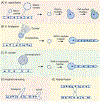
Key Figure 1.. Shape shifting fungal pathogens.
Morphology changes seen for some of the human fungal pathogens known to cause lethal systemic disease. Morphological forms on the left correspond to those seen in the environment (or at room temperature in the lab). The top four species all undergo a morphological shift to form a type of small spore that can be aerosolized and inhaled deeply into the lung. The spore germinates in the host to shape shift again into the forms shown on the right. C. albicans is a commensal organism that is commonly found growing on human skin or mucosa, so it does not require a shift to a spore form to infect a host. P. jirovecii forms cysts with sexual spores, but it is not clear they play a role in the spread of infection to new hosts. The environmental form of P. jirovecii is not known since it cannot be grown in vitro, and it is not known if there is an environmental reservoir.
Figure 2.
Figure 2.. Cryptococcus neoformans undergoes changes in size and outer capsule formation.
(A) Typical sequence of morphological changes during the budding mode of replication seen for C. neoformans. (B) Cells of opposite mating type (a and α) undergo mating to create a diploid cell, which then undergoes meiosis to create haploid nuclei that are each encased in a spore [8]. Haploid spores of the α mating type account for more than 99% of human infections. (C) Spores inhaled into a host shift back to a budding mode of replication. A thick polysaccharide capsule forms to protect the cell against stress, and large cells form which are resistant to phagocytosis by immune cells.
Figure 3.
Figure 3.. Molds in the environment produce spores that shift back to filamentous growth forms in the host.
(A-C) Molds growing as filamentous hyphae in the environment undergo morphological changes to produce distinct types of spore-forming structures. The small spores are aerosolized and then inhaled into the lung. (A) H. capsulatum produces micro- and macroconidia, which differ in size and surface structure. The smaller microconidia are thought to be more easily inhaled deep into the lung where they shift to budding to initiate an infection. Immunogenic surface molecules are cloaked, such as by the production of an outer layer of a polymer of the sugar α-glucan (green). (B) A. fumigatus produces conidia that shift back to forming septate hyphae in the host. (C) R. oryzae spores revert back to growing as coenocytic (aseptate) hyphae in the host that produce very few septa. Filamentous growth can disseminate to form a complex interconnected mycelium by (D) hyphal branching and (E) fusion of hyphal tips.
Figure 4.
Figure 4.. Candida species form multicellular structures.
Candida species divide by budding, but under certain conditions some can form (A) pseudohyphae which are multicellular chains of created by sequential budding in the same direction. (B) C. albicans can also undergo a shift to forming true hyphae characterized by smooth parallel walls. (C) Candida species form multicellular structures to avoid attack by the immune system. As described in the text, C. albicans can form multicellular hyphae and pseudohyphae that are too large to be phagocytosed. Other species that more rarely form hyphae or pseudohyphae often form cell aggregates in the host or biofilms surrounded by a matrix that resist attack by the immune system. C. albicans is also very good at forming biofilms.
Figure 5.
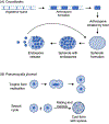
Figure 5.. Coccidioides and Pneumocystis form spores in the host.
Although many fungal species form spores in the environment, two human pathogens are worth noting for their ability to form a type of spore in the host. (A) Coccidioides species (immitis and posadasii) convert hyphal cells to arthrospores in the environment. Inhaled arthrospores shift to forming large spherules containing hundreds of endospores. Rupture of the mature spherules releases the endospores, which go on to form more spherules. (B) Haploid cells of the trophic form of P. jirovecii divide by fission. However, mating can occur between opposite mating type followed by meiosis to create a cyst form with 8 haploid nuclei.

References

    1. Brown GD et al. (2012) Hidden killers: human fungal infections. Sci Transl Med 4 (165), 165rv13. - PubMed
    1. Gow NA et al. (2002) Fungal morphogenesis and host invasion. Curr Opin Microbiol 5 (4), 366–71. - PubMed
    1. Li Z and Nielsen K (2017) Morphology Changes in Human Fungal Pathogens upon Interaction with the Host. J Fungi (Basel) 3 (4), pii: 66. doi: 10.3390/jof3040066. - DOI - PMC - PubMed
    1. Kohler JR et al. (2017) Fungi that Infect Humans. Microbiol Spectr 5 (3). - PMC - PubMed
    1. Erwig LP and Gow NA (2016) Interactions of fungal pathogens with phagocytes. Nat Rev Microbiol 14 (3), 163–76. - PubMed

Publication types

Substances