

Infectivity of human coronavirus in the brain
- PMID: 32474399
- PMCID: PMC7255711
- DOI: 10.1016/j.ebiom.2020.102799
Infectivity of human coronavirus in the brain
Abstract
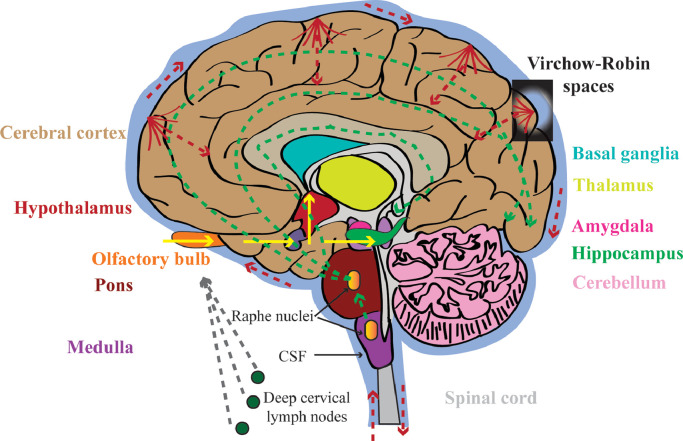
A new strain of human coronaviruses (hCoVs), Severe Acute Respiratory Syndrome Coronavirus-2 (SARS-CoV-2), has been identified to be responsible for the current outbreak of the coronavirus disease 2019 (COVID-19). Though major symptoms are primarily generated from the respiratory system, neurological symptoms are being reported in some of the confirmed cases, raising concerns of its potential for intracranial invasion and neurological manifestations, both in the acute phase and in the long-term. At present, it remains unclear the extent to which SARS-CoV-2 is present in the brain, and if so, its pathogenic role in the central nervous system (CNS). Evidence for neuroinvasion and neurovirulence of hCoVs has been recognised in animal and human studies. Given that SARS-CoV-2 belongs to the same family and shares characteristics in terms of receptor binding properties, it is worthwhile exploring its potential CNS manifestations. This review summarises previous findings from hCoVs in relation to the CNS, and compares these with the new strain, aiming to provide a better understanding of the effects of SARS-CoV-2 on the CNS.
Keywords: Brain; Coronavirus; Human; Neuroinvasion; Neurological manifestation; SARS-CoV-2.
Copyright © 2020 The Author(s). Published by Elsevier B.V. All rights reserved.
Conflict of interest statement
Declaration of Competing Interest We declare no conflicts of interest.
Figures
References
Publication types
MeSH terms
Substances
LinkOut - more resources
Full Text Sources
Other Literature Sources
Miscellaneous
