Review
. 2020 May;8(1):e000348.
doi: 10.1136/jitc-2019-000348.

Biomarkers for immunotherapy for treatment of glioblastoma

Affiliations
Review

Biomarkers for immunotherapy for treatment of glioblastoma

John P Lynes et al. J Immunother Cancer. 2020 May.

Abstract

Immunotherapy is a promising new therapeutic field that has demonstrated significant benefits in many solid-tumor malignancies, such as metastatic melanoma and non-small cell lung cancer. However, only a subset of these patients responds to treatment. Glioblastoma (GBM) is the most common malignant primary brain tumor with a poor prognosis of 14.6 months and few treatment advancements over the last 10 years. There are many clinical trials testing immune therapies in GBM, but patient responses in these studies have been highly variable and a definitive benefit has yet to be identified. Biomarkers are used to quantify normal physiology and physiological response to therapies. When extensively characterized and vigorously validated, they have the potential to delineate responders from non-responders for patients treated with immunotherapy in malignancies outside of the central nervous system (CNS) as well as GBM. Due to the challenges of current modalities of radiographic diagnosis and disease monitoring, identification of new predictive and prognostic biomarkers to gauge response to immune therapy for patients with GBM will be critical in the precise treatment of this highly heterogenous disease. This review will explore the current and future strategies for the identification of potential biomarkers in the field of immunotherapy for GBM, as well as highlight major challenges of adapting immune therapy for CNS malignancies.

Keywords: neuroimmunology; neuropathy; neurosurgery; tissue typing; tumors.


Conflict of interest statement

Competing interests: None declared.

Figures
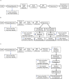
Figure 1
Figure 1
Different biomarker acquisition timepoints. (A) Standard. The current standard treatment course for patients with glioblastoma (GBM) used by practicing physicians. With this standard approach, biomarkers are only acquired from the primary resection tumor sample and usually without additional tissue samples taken during any subsequent trials. (B) Randomized neoadjuvant vs adjuvant immune therapy at recurrence. This schematic describes a more innovative approach to clinical trial design practiced by a few physicians allowing for more timepoints to acquire biomarkers. Following standard treatment, recurrence is suspected from MR imaging and patients can be enrolled on a trial to be treated with neoadjuvant immunotherapy prior to resection of the recurrent tumor or adjuvant immunotherapy after the second resection. Here, biomarkers can be collected from the primary tumor (I) and from recurrent tumor tissue following ±neoadjuvant immunotherapy (II). Additionally, non-surgical biomarkers can be collected during follow-up adjuvant immunotherapy visits (III). (C) Proposed trial scheme to maximize biomarker identification. The proposed ideal approach to designing clinical trials to ensure physicians obtain biomarkers at all critical timepoints. First, biomarkers are collected from the primary tumor sample from the initial biopsy and/or resection (I). On suspected recurrence on MRI, a second biopsy will be performed to confirm recurrence vs pseudoprogression and to collect biomarkers of the recurrent tumor prior to any immunotherapy treatment (II). Third, biomarkers will be collected from resected tumor tissue after neoadjuvant administration of immunotherapy (III). Fourth, during adjuvant immunotherapy treatment, non-surgical biomarkers can be acquired with patient follow ups (IV). Lastly, should a patient present with progression vs pseudoprogression again, biomarkers can be collected during a therapeutic surgical intervention (V).
Figure 2
Figure 2
Immunotherapy trial design to find new predictive biomarkers. (A) A flow chart detailing how retrospective clinical trials can identify predictive biomarkers. As patients enroll in immunotherapy clinical trials, researchers can retrospectively delineate the responders to treatment from the non-responders and determine biomarker differences (mutation vs wild type, presence vs absence, high vs low expression). (B) Description of how identified predictive biomarkers in retrospective studies can be rigorously tested in prospective studies. On enrollment, patients will have their tissue sampled to identify changes in biomarkers identified in the cognate retrospective study. Then they will be stratified according to these criteria and administered treatment. Response from both groups will be compared to determine if the identified biomarkers are truly predictive of response to treatment.
Figure 3
Figure 3
Trial scheme for the use of previously identified predictive biomarkers. A flow chart depicting how predictive biomarkers can be used in current clinical practices. When patients are initially diagnosed with glioblastoma (GBM), their tissue will be sampled to determine the presence of predictive biomarkers. Depending on the biomarkers present, patients will be enrolled in clinical trials that have shown response to treatment when that biomarker is present. If the patient is responding to treatment they will remain on the clinical trial, but if not, they can be switched to a different clinical trial to continue a different immune therapy treatment.
Figure 4
Figure 4
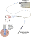
Cytokine microdialysis catheter mechanism. A diagram describing the mechanism of the microdialysis catheter. Artificial cerebrospinal fluid (CSF) perfusion fluid is pushed through the catheter using a microdialysis pump. When the fluid reaches the catheter tip, solutes are exchanged between the artificial CSF perfusion fluid and the interstitial fluid of the brain through a semi-permeable membrane. The resulting mixture is then pushed into a microvial where it can be collected and analyzed.

Similar articles

Cited by

References

    1. Lynes J, Sanchez V, Dominah G, et al. Current options and future directions in immune therapy for glioblastoma. Front Oncol 2018;8:578 10.3389/fonc.2018.00578 - DOI - PMC - PubMed
    1. Larkin J, Chiarion-Sileni V, Gonzalez R, et al. Combined nivolumab and ipilimumab or monotherapy in untreated melanoma. N Engl J Med Overseas Ed 2015;373:23–34. 10.1056/NEJMoa1504030 - DOI - PMC - PubMed
    1. Garon EB, Rizvi NA, Hui R, et al. Pembrolizumab for the treatment of Non–Small-Cell lung cancer. N Engl J Med 2015;372:2018–28. 10.1056/NEJMoa1501824 - DOI - PubMed
    1. Sanders S, Stewart DA. Targeting non-Hodgkin lymphoma with blinatumomab. Expert Opin Biol Ther 2017;17:1013–7. 10.1080/14712598.2017.1334053 - DOI - PubMed
    1. Enblad G, Karlsson H, Loskog ASI. Car T-cell therapy: the role of physical barriers and immunosuppression in lymphoma. Hum Gene Ther 2015;26:498–505. 10.1089/hum.2015.054 - DOI - PMC - PubMed

Publication types