

Radiographic findings in 240 patients with COVID-19 pneumonia: time-dependence after the onset of symptoms
- PMID: 32474630
- PMCID: PMC7260475
- DOI: 10.1007/s00330-020-06967-7
Radiographic findings in 240 patients with COVID-19 pneumonia: time-dependence after the onset of symptoms
Abstract
Objective: To analyze the most frequent radiographic features of COVID-19 pneumonia and assess the effectiveness of chest X-ray (CXR) in detecting pulmonary alterations.
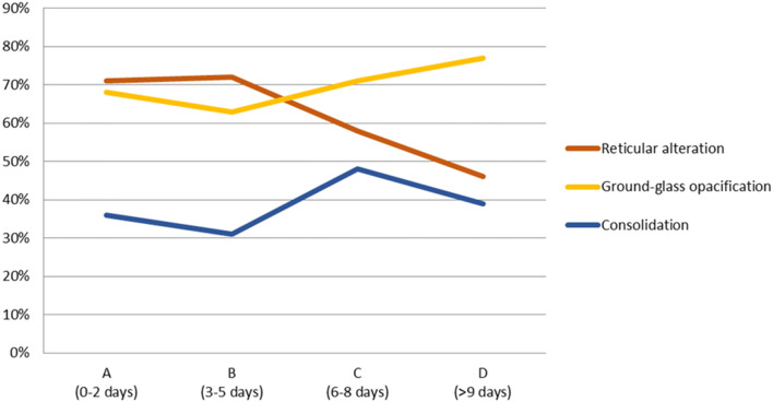
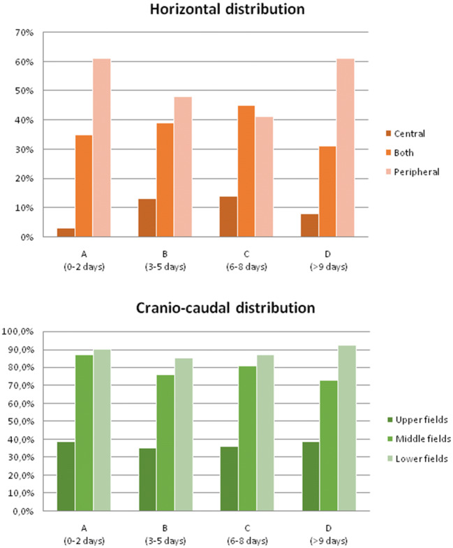
Materials and methods: CXR of 240 symptomatic patients (70% male, mean age 65 ± 16 years), with SARS-CoV-2 infection confirmed by RT-PCR, was retrospectively evaluated. Patients were clustered in four groups based on the number of days between symptom onset and CXR: group A (0-2 days), 49 patients; group B (3-5), 75 patients; group C (6-9), 85 patients; and group D (> 9), 31 patients. Alteration's type (reticular/ground-glass opacity (GGO)/consolidation) and distribution (bilateral/unilateral, upper/middle/lower fields, peripheral/central) were noted. Statistical significance was tested using chi-square test.
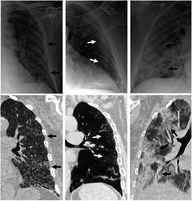
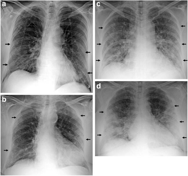
Results: Among 240 patients who underwent CXR, 180 (75%) showed alterations (group A, 63.3%; group B, 72%; group C, 81.2%; group D, 83.9%). GGO was observed in 124/180 patients (68.8%), reticular alteration in 113/180 (62.7%), and consolidation in 71/180 (39.4%). Consolidation was significantly less frequent (p < 0.01). Distribution among groups was as follows: reticular alteration (group A, 70.9%; group B, 72.2%; group C, 57.9%; group D, 46.1%), GGO (group A, 67.7%; group B, 62.9%; group C, 71%; group D, 76.9%), and consolidation (group A, 35.5%; group B, 31.4%; group C, 47.8%; group D, 38.5%). Alterations were bilateral in 73.3%. Upper, middle, and lower fields were involved in 36.7%, 79.4%, and 87.8%, respectively. Lesions were peripheral in 49.4%, central in 11.1%, or both in 39.4%. Upper fields and central zones were significantly less involved (p < 0.01).
Conclusions: The most frequent lesions in COVID-19 patients were GGO (intermediate/late phase) and reticular alteration (early phase) while consolidation gradually increased over time. The most frequent distribution was bilateral, peripheral, and with middle/lower predominance. Overall rate of negative CXR was 25%, which progressively decreased over time.
Key points: • The predominant lung changes were GGO and reticular alteration, while consolidation was less frequent. • The typical distribution pattern was bilateral, peripheral, or both peripheral and central and involved predominantly the lower and middle fields. • Chest radiography showed lung abnormalities in 75% of patients with confirmed SARS-CoV-2 infection, range varied from 63.3 to 83.9%, respectively, at 0-2 days and > 9 days from the onset of symptoms.
Keywords: COVID-19; Pneumonia; Radiography; Severe acute respiratory syndrome coronavirus 2; Thorax.
Conflict of interest statement
CB is a consultant for Bracco Imaging Italia and Doc. Congress. The other authors have nothing to disclose.
Figures
References
-
- World Health Organization . WHO Director-General’s opening remarks at the media briefing on COVID-19. Geneva: World Health Organization; 2020.
-
- World Health Organization . Coronavirus disease (COVID-19) technical guidance: laboratory testing for 2019-nCoV in humans. Geneva: World Health Organization; 2020.
MeSH terms
LinkOut - more resources
Full Text Sources
Miscellaneous
