

CT in coronavirus disease 2019 (COVID-19): a systematic review of chest CT findings in 4410 adult patients
- PMID: 32474632
- PMCID: PMC7261039
- DOI: 10.1007/s00330-020-06975-7
CT in coronavirus disease 2019 (COVID-19): a systematic review of chest CT findings in 4410 adult patients
Abstract
Objective: The objective of this systematic review was to evaluate the key imaging manifestations of COVID-19 on chest CT in adult patients by providing a comprehensive review of the published literature.
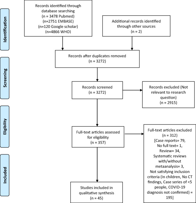
Methods: We performed a systematic literature search from the PubMed, Google Scholar, Embase, and WHO databases for studies mentioning the chest CT imaging findings of adult COVID-19 patients.
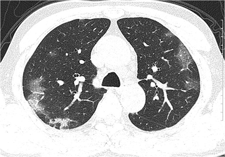
Results: A total of 45 studies comprising 4410 patients were included. Ground glass opacities (GGO), in isolation (50.2%) or coexisting with consolidations (44.2%), were the most common lesions. Distribution of GGOs was most commonly bilateral, peripheral/subpleural, and posterior with predilection for lower lobes. Common ancillary findings included pulmonary vascular enlargement (64%), intralobular septal thickening (60%), adjacent pleural thickening (41.7%), air bronchograms (41.2%), subpleural lines, crazy paving, bronchus distortion, bronchiectasis, and interlobular septal thickening. CT in early follow-up period generally showed an increase in size, number, and density of GGOs, with progression into mixed areas of GGOs plus consolidations and crazy paving, peaking at 10-11 days, before gradually resolving or persisting as patchy fibrosis. While younger adults more commonly had GGOs, extensive/multilobar involvement with consolidations was prevalent in the older population and those with severe disease.
Conclusion: This review describes the imaging features for diagnosis, stratification, and follow-up of COVID-19 patients. The most common CT manifestations are bilateral, peripheral/subpleural, posterior GGOs with or without consolidations with a lower lobe predominance. It is pertinent to be familiar with the various imaging findings to positively impact the management of these patients.
Key points: • Ground glass opacities (GGOs), whether isolated or coexisting with consolidations, in bilateral and subpleural distribution, are the most prevalent chest CT findings in adult COVID-19 patients. • Follow-up CT shows a progression of GGOs into a mixed pattern, reaching a peak at 10-11 days, before gradually resolving or persisting as patchy fibrosis. • Younger people tend to have more GGOs. Older or sicker people tend to have more extensive involvement with consolidations.
Keywords: COVID-19; Diagnostic imaging; Radiology.
Conflict of interest statement
The authors of this manuscript declare no relationships with any companies whose products or services may be related to the subject matter of the article.
Figures
References
-
- Novel coronavirus (2019-nCoV) situation reports. [cited 2020 Apr 8]. Available from: https://www.who.int/emergencies/diseases/novel-coronavirus-2019/situatio...
Publication types
MeSH terms
LinkOut - more resources
Full Text Sources
Medical
