. 2021 Jul;39(11):4175-4184.
doi: 10.1080/07391102.2020.1775123. Epub 2020 Jun 9.

Antiviral effects of probiotic metabolites on COVID-19

Affiliations

Antiviral effects of probiotic metabolites on COVID-19

Firoz Anwar et al. J Biomol Struct Dyn. 2021 Jul.

Abstract

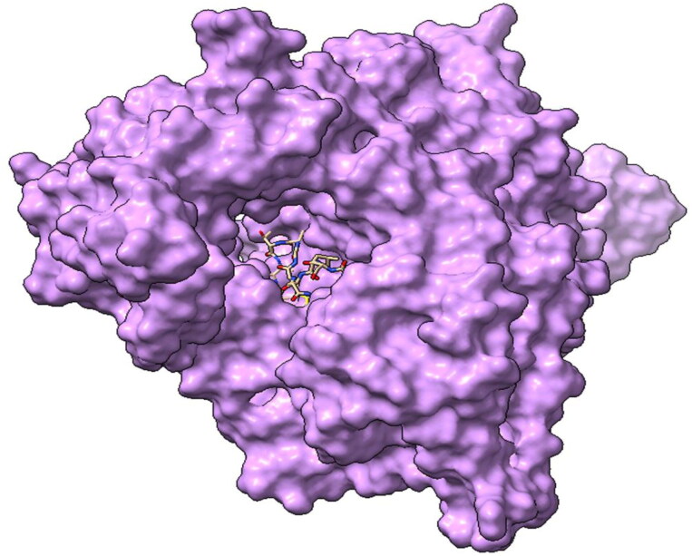
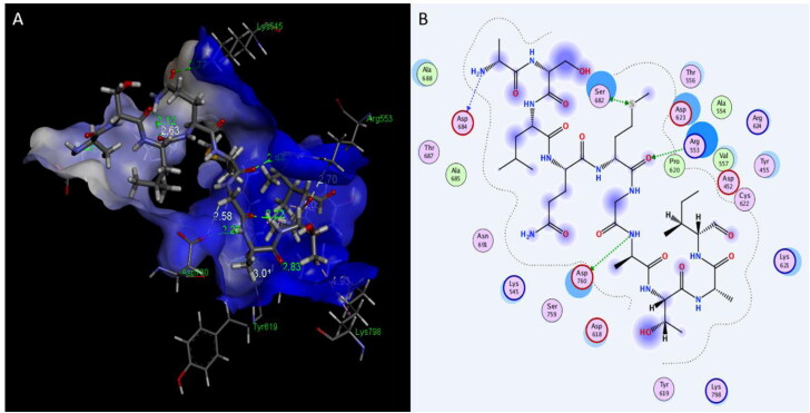
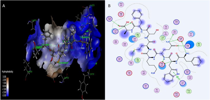
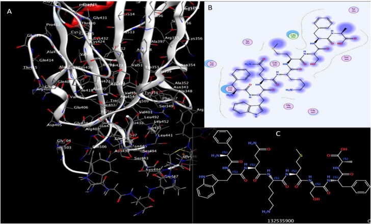
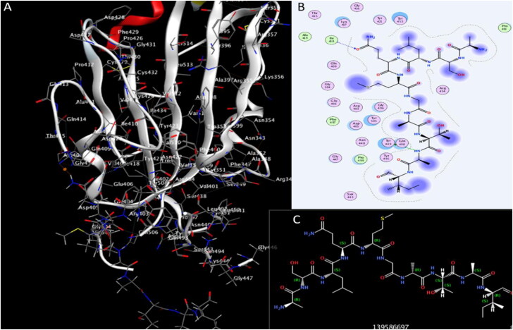
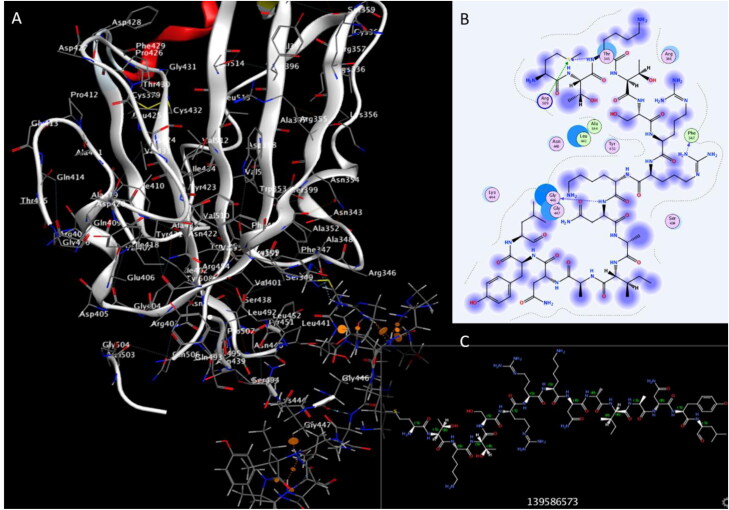
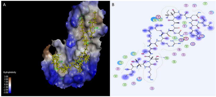
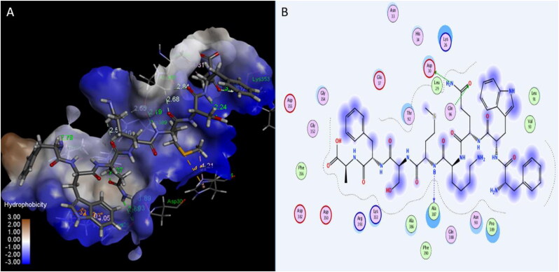
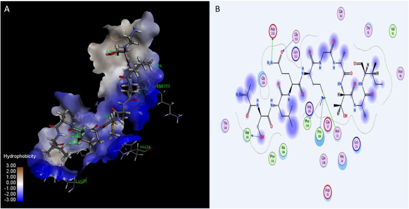
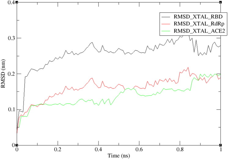
SARS coronavirus (COVID-19) is a real health challenge of the 21st century for scientists, health workers, politicians, and all humans that has severe cause epidemic worldwide. The virus exerts its pathogenic activity through by mechanism and gains the entry via spike proteins (S) and Angiotensin-Converting Enzyme 2 (ACE2) receptor proteins on host cells. The present work is an effort for a computational target to block the residual binding protein (RBP) on spike proteins (S), Angiotensin-Converting Enzyme 2 (ACE2) receptor proteins by probiotics namely Plantaricin BN, Plantaricin JLA-9, Plantaricin W, Plantaricin D along with RNA-dependent RNA polymerase (RdRp). Docking studies were designed in order to obtain the binding energies for Plantaricin metabolites. The binding energies for Plantaricin W were -14.64, -11.1 and -12.68 for polymerase, RBD and ACE2 respectively comparatively very high with other compounds. Plantaricin W, D, and JLA-9 were able to block the residues (THR556, ALA558) surrounding the deep grove catalytic site (VAL557) of RdRp making them more therapeutically active for COVID-19. Molecular dynamics studies further strengthen stability of the complexes of plantaricin w and SARS-CoV-2 RdRp enzyme, RBD of spike protein, and human ACE2 receptor. The present study present multi-way options either by blocking RBD on S proteins or interaction of S protein with ACE2 receptor proteins or inhibiting RdRp to counter any effect of COVID-19 by Plantaricin molecules paving a way that can be useful in the treatment of COVID-19 until some better option will be available.Communicated by Ramaswamy H. Sarma.

Keywords: ACE2; COVID-19; RNA dependent RNA polymerase; plantaricin; probiotics; spike proteins.


Figures

Figure 1.
Figure 1.
Three-dimensional structure of RNA-dependent-RNA polymerase (RdRp) enzymes with Plantaricin W blocking the active site cavity.
Figure 2.
Figure 2.
Interaction of Plantaricin D and RNA-dependent-RNA polymerase(RdRp) enzymes. (A) The three-dimensional structure of ligand is surrounded d by the hydrophobic surface of RdRp. (B) Two-dimensional interaction shows hydrogen bonds in green dotted lines.
Figure 3.
Figure 3.
Interaction of Plantaricin JLA and RNA-dependent-RNA polymerase (RdRp) enzymes. (A) The three-dimensional structure of ligand is surrounded d by the hydrophobic surface of RdRp. (B) Two-dimensional interaction shows hydrogen bonds in green dotted lines.
Figure 4.
Figure 4.
Interaction of Plantaricin JLA and SARS-CoV-2 receptor-binding domain (RBD). (A) The three-dimensional structure of ligand and amino acids, the RBD is coloured white. (B) Two-dimensional interaction shows hydrogen bonds in green dotted lines. (C) Two-dimensional structure and PubChem ID of Plantaricin JLA ligand.
Figure 5.
Figure 5.
Interaction of Plantaricin D and SARS-CoV-2 receptor-binding domain (RBD). (A) The three-dimensional structure of ligand and amino acids, the RBD is coloured white. (B) Two-dimensional interaction shows hydrogen bonds in green dotted lines. (C) Two-dimensional structure and PubChem ID of Plantaricin D ligand.
Figure 6.
Figure 6.
Interaction of Plantaricin W and SARS-CoV-2 receptor-binding domain (RBD). (A) The three-dimensional structure of ligand and amino acids, the RBD is coloured white. (B) Two-dimensional interaction shows hydrogen bonds in green dotted lines. (C) Two-dimensional structure and PubChem ID of Plantaricin W ligand.
Figure 7.
Figure 7.
Interaction of Plantaricin W and angiotensin-converting enzyme 2 (ACE2) receptor. (A) The three-dimensional structure of ligand is surrounded d by the hydrophobic surface of ACE2. (B) Two-dimensional interaction shows hydrogen bonds in green dotted lines.
Figure 8.
Figure 8.
Interaction of Plantaricin JLA-9 and angiotensin-converting enzyme 2 (ACE2) receptor. (A) The three-dimensional structure of ligand is surrounded d by the hydrophobic surface of ACE2 (B) Two-dimensional interaction shows hydrogen bonds in green dotted lines.
Figure 9.
Figure 9.
Interaction of Plantaricin D and angiotensin-converting enzyme 2 (ACE2) receptor. (A) The three-dimensional structure of ligand is surrounded d by the hydrophobic surface of ACE2. (B) Two-dimensional interaction shows hydrogen bonds in green dotted lines.
Figure 10.
Figure 10.
RMSD plot of RBD, RdRp, and ACE2 proteins backbones in complex with plantaricin w, over one nanosecond period.

References

    1. Aanouz I., Belhassan A., El Khatabi K., Lakhlifi T., El Idrissi M., & Bouachrine M. (2020). Moroccan medicinal plants as inhibitors of COVID-19: Computational investigations. Journal of Biomolecular Structure & Dynamics, 6, 1–9. 10.1080/07391102.2020.1758790 - DOI - PMC - PubMed
    1. Al Kassaa I. (2016). New insights on antiviral probiotics: From research to applications. New Insights on Antiviral Probiotics: From Research to Applications. 10.1007/978-3-319-49688-7 - DOI
    1. Albarracin L., Kobayashi H., Iida H., Sato N., Nochi T., Aso H., Salva S., Alvarez S., Kitazawa H., & Villena J. (2017). Transcriptomic analysis of the innate antiviral immune response in porcine intestinal epithelial cells: Influence of immunobiotic lactobacilli. Frontiers in Immunology, 8, 57 10.3389/fimmu.2017.00057 - DOI - PMC - PubMed
    1. Berman H. M., Battistuz T., Bhat T. N., Bluhm W. F., Bourne P. E., Burkhardt K., … Zardecki C. (2002). The protein data bank. Acta Crystallographica Section D: Biological Crystallography. 10.1107/S0907444902003451 - DOI - PubMed
    1. Boopathi S., Poma A. B., & Kolandaivel P. (2020). Novel 2019 Coronavirus structure, mechanism of action, antiviral drug promises and rule out against its treatment. Journal of Biomolecular Structure & Dynamics, 30, 1–10. 10.1080/07391102.2020.1758788 - DOI - PMC - PubMed

Substances