. 2020 Jun;53(6):329-334.
doi: 10.5483/BMBRep.2020.53.6.065.

The activation of NLRP3 inflammasome potentiates the immunomodulatory abilities of mesenchymal stem cells in a murine colitis model

Affiliations

The activation of NLRP3 inflammasome potentiates the immunomodulatory abilities of mesenchymal stem cells in a murine colitis model

Ji-Su Ahn et al. BMB Rep. 2020 Jun.

Abstract

Inflammasomes are cytosolic, multiprotein complexes that act at the frontline of the immune responses by recognizing pathogen- or danger-associated molecular patterns or abnormal host molecules. Mesenchymal stem cells (MSCs) have been reported to possess multipotency to differentiate into various cell types and immunoregulatory effects. In this study, we investigated the expression and functional regulation of NLR Family Pyrin Domain Containing 3 (NLRP3) inflammasome in human umbilical cord blood-derived MSCs (hUCB-MSCs). hUCB-MSCs expressed inflammasome components that are necessary for its complex assembly. Interestingly, NLRP3 inflammasome activation suppressed the differentiation of hUCB-MSCs into osteoblasts, which was restored when the expression of adaptor proteins for inflammasome assembly was inhibited. Moreover, the suppressive effects of MSCs on T cell responses and the macrophage activation were augmented in response to NLRP3 activation. In vivo studies using colitic mice revealed that the protective abilities of hUCB-MSCs increased after NLRP3 stimulation. In conclusion, our findings suggest that the NLRP3 inflammasome components are expressed in hUCB-MSCs and its activation can regulate the differentiation capability and the immunomodulatory effects of hUCB-MSCs. [BMB Reports 2020; 53(6): 329-334].


Conflict of interest statement

CONFLICTS OF INTEREST

The authors have no conflicting interests.

Figures

Fig. 1
Fig. 1
The expression of inflammasome components and viability, proliferation, and death of hUCB-MSCs after NLRP3 inflammasome activation. (A) Expressions of NLRP3, ASC, pro or cleaved caspase-1, pro or mature IL-1β were analyzed by immunoblotting in hUCB-MSCs after treatment with LPS, ATP or LPS+ATP. (B) The concentration of mature IL-1β in the culture supernatant of hUCB-MSCs was measured by ELISA. (C) Cell viability was detected by CCK-8. (D) Cell proliferation was determined by CPDL analysis. (E) Cell death was assessed by Live/Dead staining. Bar, 500 μm. Results show 1 representative experiment of at least 3 independent experiments. Results are shown as mean ± SD.
Fig. 2
Fig. 2
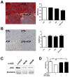
Osteogenic and adipogenic differentiation of NLRP3-activated hUCB-MSCs. (A) hUCB-MSCs were cultured in osteoblast induction media and stained with Alizarin Red S. Stain was eluted and the absorbance was measured at 570 nm. Bar, 250 μm. (B) hUCB-MSCs were differentiated into adipocytes and stained by Oil Red O. Stain was eluted and the absorbance was measured at 500 nm. Bar, 250 μm. (C) siRNA for ASC was transfected to hUCB-MSCs and a decrease in protein level was observed. (D) siRNA-treated hUCB-MSCs were induced for osteogenic differentiation. The eluted stain was quantified. Results show 1 representative experiment of at least 3 independent experiments. Results are shown as mean ± SD. **P < .01, ***P < .001.
Fig. 3
Fig. 3
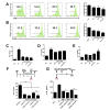
Immunoregulatory properties of NLRP3-stimulated hUCB-MSCs. (A, B) PBMCs were stained with CFSE and then activated by (A) concanavalin A or (B) anti-CD3/28 to induce the proliferation of pan leukocytes or T cells. (C-E) CD4+ T cells isolated from PBMCs were co-cultured with hUCB-MSCs. (C, D) Th1 and Th2 differentiation was induced in the presence of hUCB-MSCs. (C) IFN-g and (D) IL-4 productions were measured. (E) Naïve CD4+ T cells were co-cultured with hUCB-MSCs and IL-10 production was measured. (F) THP-1-derived M1 macrophages were co-cultured with hUCB-MSCs. TNF-α concentration in the culture media was detected. (G) NLRP3-activated THP-1-derived macrophages were co-cultured with hUCB-MSCs. IL-1β level in the culture media was detected. Results show 1 representative experiment of at least 3 independent experiments. Results are shown as mean ± SD. *P < .05, **P < .01, ***P < .001.
Fig. 4
Fig. 4
Protective efficacy of NLRP3-stimulated hUCB-MSCs in the DSS-induced colitis model. DSS-induced colitic mice were injected with hUCB-MSCs. (A) Mice were monitored for survival rate and body weight loss. The statistical significance was calculated by comparing other groups against 'DSS+PBS' group (marked as '+'). (B) On day 10, the disease activity index was determined. (C) After sacrifice on day 12, colon length was measured. (D) Colon sections were prepared and stained with H&E. Histopathologic evaluation was performed by determining the destruction of epithelial structure and lymphocyte infiltration. Bar, 500 μm. Results are shown as mean ± SD. *P < .05, **P < .01, ***P < .001.

References

    1. Akira S, Uematsu S, Takeuchi O. Pathogen recognition and innate immunity. Cell. 2006;124:783–801. doi: 10.1016/j.cell.2006.02.015. - DOI - PubMed
    1. Beutler B, Eidenschenk C, Crozat K, et al. Genetic analysis of resistance to viral infection. Nat Rev Immunol. 2007;7:753–766. doi: 10.1038/nri2174. - DOI - PubMed
    1. Hoffmann JA. The immune response of Drosophila. Nature. 2003;426:33–38. doi: 10.1038/nature02021. - DOI - PubMed
    1. Medzhitov R. Recognition of microorganisms and activation of the immune response. Nature. 2007;449:819–826. doi: 10.1038/nature06246. - DOI - PubMed
    1. Kato H, Takahasi K, Fujita T. RIG-I-like receptors: cytoplasmic sensors for non-self RNA. Immunol Rev. 2011;243:91–98. doi: 10.1111/j.1600-065X.2011.01052.x. - DOI - PubMed