Review
. 2020 Jun;39(26):4909-4924.
doi: 10.1038/s41388-020-1336-y. Epub 2020 May 31.

PROteolysis TArgeting Chimeras (PROTACs) as emerging anticancer therapeutics

Affiliations
Review

PROteolysis TArgeting Chimeras (PROTACs) as emerging anticancer therapeutics

Sajid Khan et al. Oncogene. 2020 Jun.

Abstract

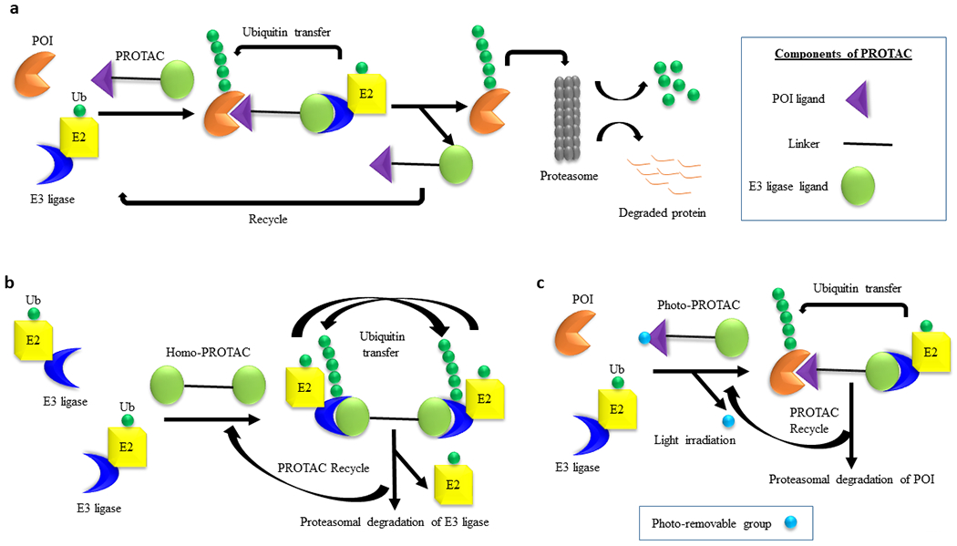
Using PROteolysis TArgeting Chimeras (PROTACs) to degrade proteins that are important for tumorigenesis has emerged as a potential therapeutic strategy for cancer. PROTACs are heterobifunctional molecules consisting of one ligand for binding to a protein of interest (POI) and another to an E3 ubiquitin (E3) ligase, connected via a linker. PROTACs recruit the E3 ligase to the POI and cause proximity-induced ubiquitination and degradation of the POI by the ubiquitin-proteasome system (UPS). PROTACs have been developed to degrade a variety of cancer targets with unprecedented efficacy against a multitude of tumor types. To date, most of the PROTACs developed have utilized ligands to recruit E3 ligases that are ubiquitously expressed in both tumor and normal tissues. These PROTACs can cause on-target toxicities if the POIs are not tumor-specific. Therefore, identifying and recruiting the E3 ligases that are enriched in tumors with minimal expression in normal tissues holds the potential to develop tumor-specific/selective PROTACs. In this review, we will discuss the potential of PROTACs to become anticancer therapeutics, chemical and bioinformatics approaches for PROTAC design, and safety concerns with a special focus on the development of tumor-specific/selective PROTACs. In addition, the identification of tumor types in terms of solid versus hematological malignancies that can be best targeted with PROTAC approach will be briefly discussed.


Conflict of interest statement

CONFLICT OF INTEREST: S.K., Y.H., X.Z, G.Z., and D.Z. are inventors of two pending patent applications for use of Bcl-xL PROTACs as senolytic and antitumor agents. G.Z., and D.Z. are co-founders of and have equity in Dialectic Therapeutics, which develops Bcl-xL PROTACs to treat cancer. The remaining authors declare no competing financial interests.

Figures

Fig. 1.
Fig. 1.. Mechanisms of PROTAC-mediated protein degradation.
a, Schematic representation of the general mechanism of PROTAC-induced degradation of POI. A PROTAC molecule recruits an E3 ligase to a POI followed by polyubiquitin of the latter by E2 conjugating enzyme. The polyubiquitinated POI is recognized by the proteasome for its degradation. The PROTAC molecule is recycled to induce next round of degradation. The three different structural components of a PROTAC are depicted in the box. b, Mechanism of the self-degradation of an E3 ligase with homo-PROTACs. A homo-PROTAC recruits an E3 ligase molecule (e.g., CRBN or VHL) to another E3 ligase molecule followed by bidirectional polyubiquitination of E3 ligase molecules and their subsequent degradation by the proteasome. c, Mechanism of POI degradation by Photo-PROTACs. In a photo-PROTAC, a photo-removable group is attached to the POI ligand. Upon irradiation with external light, the photo-removable group is detached from the photo-PROTAC converting it to an active PROTAC for the degradation of POI.
Fig. 2.
Fig. 2.. Milestones in PROTAC discovery.
Numbers in superscript indicate reference number.
Fig. 3.
Fig. 3.. Profile of E3 ligase expression in normal tissues.
The original RNA-sequencing (RNA-seq) data used for the analyses were obtained from the GTEx Portal (https://www.gtexportal.org). Heatmap was drawn based on their gene expression levels (RPKM). Red circle indicates selective E3 cluster in tissues.
Fig. 4.
Fig. 4.. Distribution of E3 ligases in normal tissues.
a, Percentage of E3 ligases in normal tissues according to the classification criteria shown in Table 3. b, Immunohistochemistry (IHC) staining of representative E3 ligases in 7 normal tissues. The figures were acquired from the Human Protein Atlas (THPA) (https://www.proteinatlas.org). Red rectangle indicates high expression of E3 ligases in the tissues.
Fig. 5.
Fig. 5.. Identification of tumor-specific/selective E3 ligases.
a, Tumor-specific/selective E3 ligases in 12 tumor tissues. BLCA, bladder urothelial carcinoma; BRCA, breast invasive carcinoma; COAD, colon adenocarcinoma; HNSC, head and neck squamous cell carcinoma; KICH, kidney chromophobe; KIRC, kidney renal clear cell carcinoma; KIRP, kidney renal papillary cell carcinoma; LIHC, liver hepatocellular carcinoma; LUAD, lung adenocarcinoma; LUSC, lung squamous cell carcinoma; PRAD, prostate adenocarcinoma; THCA, thyroid carcinoma. b, BIRC7 expression (RPKM) in 12 tumor and corresponding normal tissues. Comparisons were made by two-tailed Student’s t-test.

Similar articles

Cited by

References

    1. Bai L, Zhou H, Xu R, Zhao Y, Chinnaswamy K, McEachern D et al. A Potent and Selective Small-Molecule Degrader of STAT3 Achieves Complete Tumor Regression In Vivo. Cancer Cell 2019; 36: 498–511 e417. - PMC - PubMed
    1. Zhou H, Bai L, Xu R, Zhao Y, Chen J, McEachern D et al. Structure-Based Discovery of SD-36 as a Potent, Selective, and Efficacious PROTAC Degrader of STAT3 Protein. J Med Chem 2019; 62: 11280–11300. - PMC - PubMed
    1. Zhao Q, Ren C, Liu L, Chen J, Shao Y, Sun N et al. Discovery of SIAIS178 as an Effective BCR-ABL Degrader by Recruiting Von Hippel-Lindau (VHL) E3 Ubiquitin Ligase. J Med Chem 2019; 62: 9281–9298. - PubMed
    1. Burslem GM, Smith BE, Lai AC, Jaime-Figueroa S, McQuaid DC, Bondeson DP et al. The Advantages of Targeted Protein Degradation Over Inhibition: An RTK Case Study. Cell Chem Biol 2018; 25: 67–77.e63. - PMC - PubMed
    1. Gonzalez TL, Hancock M, Sun S, Gersch CL, Larios JM, David W et al. Targeted degradation of activating estrogen receptor α ligand-binding domain mutations in human breast cancer. Breast Cancer Res Treat 2020; 180: 611–622. - PubMed

Publication types

MeSH terms