

Epidemiological investigation of porcine pseudorabies virus and its coinfection rate in Shandong Province in China from 2015 to 2018
- PMID: 32476312
- PMCID: PMC7263908
- DOI: 10.4142/jvs.2020.21.e36
Epidemiological investigation of porcine pseudorabies virus and its coinfection rate in Shandong Province in China from 2015 to 2018
Abstract
Background: Pseudorabies, also known as Aujeszky's disease, is caused by the pseudorabies virus (PRV) and has been recognized as a critical disease affecting the pig industry and a wide range of animals around the world, resulting in great economic losses each year. Shandong province, one of the most vital food animal-breeding regions in China, has a very dense pig population, within which pseudorabies infections were detected in recent years. The data, however, on PRV epidemiology and coinfection rates of PRV with other major swine diseases is sparse.
Objectives: This study aimed to investigate the PRV epidemiology in Shandong and analyze the current control measures.
Methods: In this study, a total number of 16,457 serum samples and 1,638 tissue samples, which were collected from 362 intensive pig farms (≥ 300 sows/farm) covered all cities in Shandong, were tested by performing enzyme-linked immunosorbent assay (ELISA) and polymerase chain reaction (PCR).
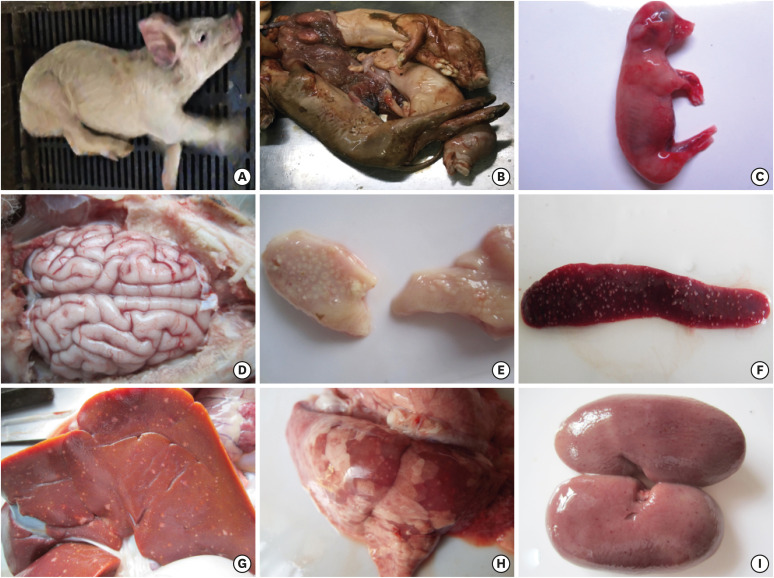
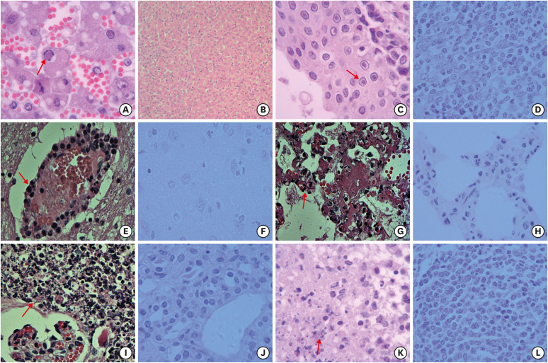
Results: Overall, 52.7% and 91.5% of the serum samples were positive for PRV-gE and -gB, respectively, based on ELISA results. In addition, 15.7% of the tissue samples were PCR positive for PRV. The coinfection rates of PRV with porcine circovirus type 2 (PCV2), porcine reproductive and respiratory syndrome virus, and classical swine fever virus were measured; coinfection with PCV2 was 35.0%, higher than those of the other two viruses. Macroscopic and microscopic lesions were observed in various tissues during histopathological examination.
Conclusions: The results demonstrate the PRV prevalence and its coinfection rates in Shandong province and indicate that pseudorabies is endemic in pig farms in this region. This study provides epidemiological data that can be useful in the prevention and control of pseudorabies in Shandong, China.
Keywords: Pseudorabies virus; coinfection; porcine circovirus; veterinary epidemiology.
© 2020 The Korean Society of Veterinary Science.
Conflict of interest statement
The authors have no conflicts of interest to declare.
Figures
Similar articles
-
Epidemiological investigation of porcine circovirus type 2 and its coinfection rate in Shandong province in China from 2015 to 2018.BMC Vet Res. 2021 Jan 7;17(1):17. doi: 10.1186/s12917-020-02718-4. BMC Vet Res. 2021. PMID: 33413367 Free PMC article.
-
Prevalence of Porcine Pseudorabies Virus and Its Coinfection Rate in Heilongjiang Province in China from 2013 to 2018.Viral Immunol. 2020 Oct;33(8):550-554. doi: 10.1089/vim.2020.0025. Epub 2020 May 12. Viral Immunol. 2020. PMID: 32397944
-
The epidemiological investigation of co-infection of major respiratory bacteria with pseudorabies virus in intensive pig farms in China.Vet Med Sci. 2021 Jan;7(1):175-183. doi: 10.1002/vms3.289. Epub 2020 Jun 24. Vet Med Sci. 2021. PMID: 32583623 Free PMC article.
-
Co-Infection of Swine with Porcine Circovirus Type 2 and Other Swine Viruses.Viruses. 2019 Feb 21;11(2):185. doi: 10.3390/v11020185. Viruses. 2019. PMID: 30795620 Free PMC article. Review.
-
The prevalence of pseudorabies virus in China from 2010 to 2024: a systematic review and meta-analysis.BMC Vet Res. 2025 Aug 12;21(1):513. doi: 10.1186/s12917-025-04924-4. BMC Vet Res. 2025. PMID: 40797260 Free PMC article.
Cited by
-
Epidemiological investigation of porcine circovirus type 2 and its coinfection rate in Shandong province in China from 2015 to 2018.BMC Vet Res. 2021 Jan 7;17(1):17. doi: 10.1186/s12917-020-02718-4. BMC Vet Res. 2021. PMID: 33413367 Free PMC article.
-
Current Status and Challenge of Pseudorabies Virus Infection in China.Virol Sin. 2021 Aug;36(4):588-607. doi: 10.1007/s12250-020-00340-0. Epub 2021 Feb 22. Virol Sin. 2021. PMID: 33616892 Free PMC article. Review.
-
The Epidemiological Analysis of Pseudorabies Virus and Pathogenicity of the Variant Strain in Shandong Province.Front Vet Sci. 2022 Mar 2;9:806824. doi: 10.3389/fvets.2022.806824. eCollection 2022. Front Vet Sci. 2022. PMID: 35310414 Free PMC article.
-
Construction of a quadruple gene-deleted vaccine confers complete protective immunity against emerging PRV variant challenge in piglets.Virol J. 2022 Jan 25;19(1):19. doi: 10.1186/s12985-022-01748-8. Virol J. 2022. PMID: 35078501 Free PMC article.
-
Construction of Two Recombinant Pseudorabies Viruses with Deletion of Virulence Genes and Evaluation of Their Immune Protection in Mice and Piglets.Vaccines (Basel). 2025 Mar 27;13(4):359. doi: 10.3390/vaccines13040359. Vaccines (Basel). 2025. PMID: 40333253 Free PMC article.
References
-
- Mettenleiter TC. Aujeszky's disease (pseudorabies) virus: the virus and molecular pathogenesis--state of the art, June 1999. Vet Res. 2000;31(1):99–115. - PubMed
-
- Crandell RA. Pseudorabies (Aujeszky's disease) Vet Clin North Am Large Anim Pract. 1982;4(2):321–331. - PubMed
-
- Rziha HJ, Mettenleiter TC, Ohlinger V, Wittmann G. Herpesvirus (pseudorabies virus) latency in swine: occurrence and physical state of viral DNA in neural tissues. Virology. 1986;155(2):600–613. - PubMed
-
- Müller T, Hahn EC, Tottewitz F, Kramer M, Klupp BG, Mettenleiter TC, Freuling C. Pseudorabies virus in wild swine: a global perspective. Arch Virol. 2011;156(10):1691–1705. - PubMed
MeSH terms
Grants and funding
LinkOut - more resources
Full Text Sources
