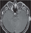
Isolated optic nerve sarcoidosis
- PMID: 32476840
- PMCID: PMC7170143
- DOI: 10.36141/svdld.v34i2.5410
Isolated optic nerve sarcoidosis
Abstract
Objective: To report three cases of sarcoidosis confined to the optic nerve. Methods: Chart review of clinical, laboratory, imaging, and optic nerve biopsy findings and a review of the literature. Results: All three cases presented with progressive visual loss and showed enhancement of the intraorbital optic nerve on magnetic resonance imaging. There was no evidence for systemic disease, including a negative workup for sarcoidosis or other infiltrative pathologies. Optic nerve biopsy in each case showed non-caseating granulomas consistent with sarcoidosis. Conclusions: Sarcoidosis confined to the optic nerve is a rare phenomenon but should still be considered in the differential diagnosis of progressive optic neuropathy, even in the absence of systemic disease. (Sarcoidosis Vasc Diffuse Lung Dis 2017; 34: 179-183).
Keywords: optic nerve; optic neuritis; optic neuropathy; sarcoidosis.
Copyright: © 2017.
Figures


References
-
- Wu JJ, Schiff KR. Am Fam Physician. 2004/08/05 ed. 2. Vol. 70. California 92103, USA: Department of Family and Preventive Medicine, University of California, San Diego, School of Medicine, La Jolla; 2004. Sarcoidosis; pp. 312–22. j1wu@ucsd.edu . - PubMed
-
- Phillips YL, Eggenberger ER. Curr Opin Ophthalmol. 2010/08/26 ed. 6. Vol. 21. USA: Department of Neurology and Ophthalmology, Michigan State University, East Lansing, Michigan; 2010. Neuro-ophthalmic sarcoidosis; pp. 423–9. - PubMed
-
- Bezo C, Majzoub S, Nochez Y, Leruez S, Charlin JF, Milea D, et al. J Fr Ophtalmol. 2013/04/03 ed. 6. Vol. 36. France: Service d’ophtalmologie, CHRU de Tours, boulevard Tonnelle, 37000 Tours; 2013. Ocular and neuro-ophthalmic manifestations of sarcoidosis: retrospective study of 30 cases; pp. 473–80. ChBezo@chu-angers.fr . - PubMed
Publication types
LinkOut - more resources
Full Text Sources
