Case Reports
. 2018;35(4):395-398.
doi: 10.36141/svdld.v35i4.7047. Epub 2020 Mar 9.

Necrotising sarcoid granulomatosis. A rare granulomatous disease

Affiliations
Case Reports

Necrotising sarcoid granulomatosis. A rare granulomatous disease

Martina Doubková et al. Sarcoidosis Vasc Diffuse Lung Dis. 2018.

Abstract

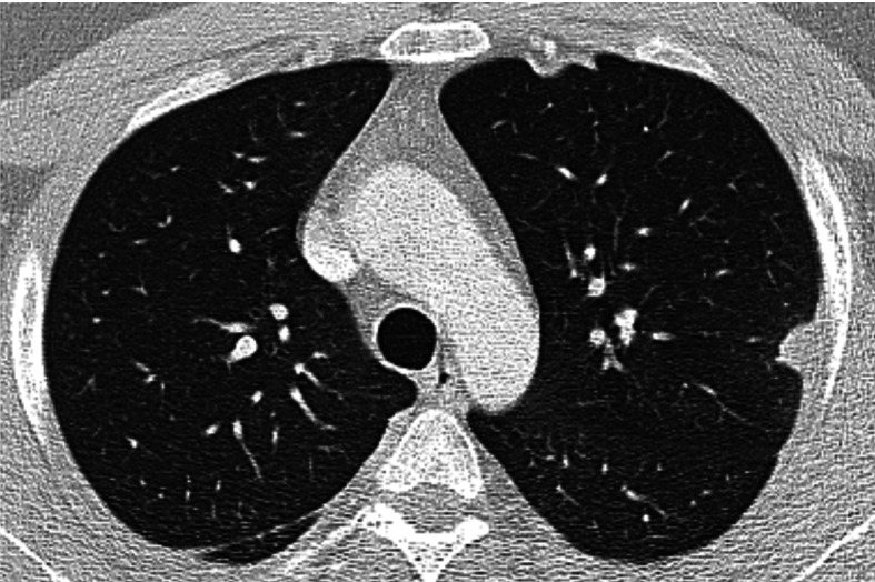
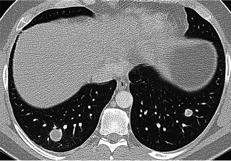
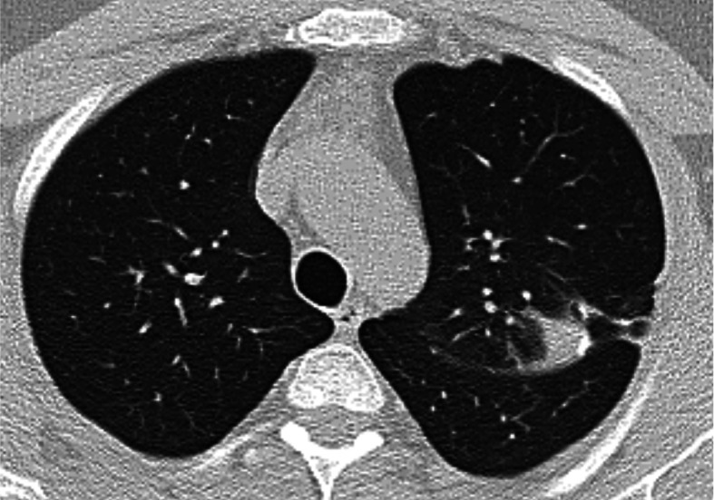
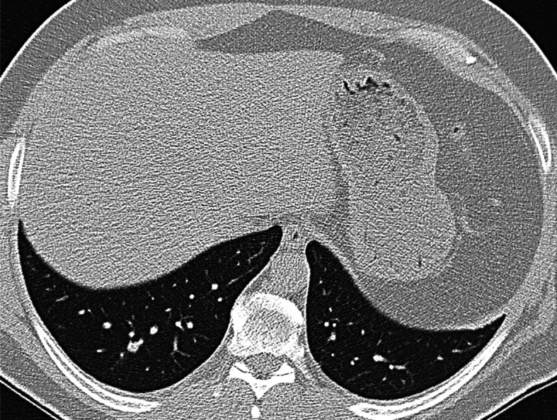
Introduction: Necrotizing sarcoid granulomatosis (NSG) is a very rare disease of unknown etiology characterized by sarcoid-like granulomas, vasculitis and necrosis in pulmonary and extrapulmonary localizations. Case report: We describe a case of a 34-year-old Caucasian male with fever, pleural pain, and nodular pulmonary opacities on chest radiograph. Histological examination of the lung tissue confirmed NSG. Diagnostically, infectious causes, vasculitis, and malignancy were excluded. A tendency to partial regression was observed, without the need for corticosteroid treatment. Conclusion: NSG is a rare disease which must be distinguished from other systemic diseases including vasculitides. The key to diagnosis, emphasized in our paper, is the histopathological finding. The course of NSG is similar to sarcoidosis. Corticosteroids are considered the treatment of choice, but the disease exhibits a tendency towards spontaneous regression. (Sarcoidosis Vasc Diffuse Lung Dis 2018; 35: 395-398).

Keywords: differential diagnosis; histopathological diagnosis; necrotising sarcoid granulomatosis.


Figures

Fig. 1.
Fig. 1.
Chest HRCT, transverse plane, initial examination. At the aortic arch level, a subpleural nodule in the apical S1+2 segments of the left upper lobe with pleural thickening in the adjacent area. Subpleural lesions are quite typical for necrotizing sarcoid granulomatosis (NSG) without predilection for upper or lower lobe involvement. In the right lung dorsally, a slight thickening of the pleura and of the major interlobar fissure
Fig. 2.
Fig. 2.
Chest HRCT, initial examination, transversal plane at the level of lower lobes shows spherical nodules with smooth edges, with no apparent spiculations extending into the surrounding pulmonary parenchyma. A larger, 18 mm nodule in the S10 segment of the right lower lobe; a smaller one, 11 mm, in the S9 segment of the left lower lobe. Among frequent findings in NSG are nodular lesions with smooth or slightly irregular borders that may exhibit cavitations
Fig. 3.
Fig. 3.
Chest HRCT, transverse plane, two-month follow-up. A tiny ‘consolidation’ of the pulmonary parenchyma with isolated calcifications and fibrous streaks extending towards the thickened pleura is present at the upper left lobe level in S1+2. The original S1+2 nodule adjacent to the pleura has partially regressed – a sign of a certain development of the disease over time
Fig. 4.
Fig. 4.
Histopathological findings. Microscopically, confluent non-caseating granulomas with extensive necrosis are seen in the lung parenchyma. The granulomas are of the sarcoid type, palisaded by Langerhans-type giant cells and mononuclear lymphohistiocytes with central necrosis. Another typical finding is the presence of vasculitis, usually granulomatous, and a large zone of necrosis. a) Large zone of necroris (×50); b) granulomatous vasculitis (×100); c) confluent granulomas and vasculitis (× 100).
Fig. 5.
Fig. 5.
Chest HRCT, transverse plane, two-month follow-up. Compared with the initial examination, multiple nodules in the pulmonary parenchyma, predominantly in subpleural localization, have partially or completely disappeared. An 8 mm nodule (previously 18 mm) is present in S10 of the left lower lobe (indicated by the arrow)

Similar articles

Cited by

References

    1. Liebow AA. The J. Burns Amberson lecture – pulmonary angiitis and granulomatosis. Am Rev Respir Dis. 1973;108(1):1–18. - PubMed
    1. Churg A. Pulmonary angiitis and granulomatosis revisited. Hum Pathol. 1983;14(10):868–883. - PubMed
    1. Churg A, Carrington GB, Gupta R. Necrotizing sarcoid granulomatosis. Chest. 1979;76(4):406–413. - PubMed
    1. Quaden C, Tillie-Leblond I, Delobbe A, et al. Necrotising sarcoid granulomatosis: clinical, functional, endoscopical and radiographical evaluations. Eur Respir J. 2005;26(5):778–85. - PubMed
    1. Bouman KP, Slabbynck H, Cuykens JJ, Galdermans D, Coolen D, Kock M. Necrotising sarcoid granulomatosis with uveitis: a variant of sarcoidosis. Acta Clin Belg. 1997;52(6):367–370. - PubMed

Publication types

LinkOut - more resources