. 2020 May 13:11:549.
doi: 10.3389/fimmu.2020.00549. eCollection 2020.

Herpes Simplex Virus 1 UL2 Inhibits the TNF-α-Mediated NF-κB Activity by Interacting With p65/p50

Affiliations

Herpes Simplex Virus 1 UL2 Inhibits the TNF-α-Mediated NF-κB Activity by Interacting With p65/p50

Mingsheng Cai et al. Front Immunol. .

Abstract

Herpes simplex virus 1 (HSV-1) is a large double-stranded DNA virus that encodes at least 80 viral proteins, many of which are involved in the virus-host interaction and are beneficial to the viral survival and reproduction. However, the biological functions of some HSV-1-encoded proteins are not fully understood. Nuclear factor κB (NF-κB) activation is the major antiviral innate response, which can be triggered by various signals induced by cellular receptors from different pathways. Here, we demonstrated that HSV-1 UL2 protein could antagonize the tumor necrosis factor α (TNF-α)-mediated NF-κB activation. Co-immunoprecipitation assays showed that UL2 could interact with the NF-κB subunits p65 and p50, which also revealed the region of amino acids 9 to 17 of UL2 could suppress the NF-κB activation and interact with p65 and p50, and UL2 bound to the immunoglobulin-like plexin transcription factor functional domain of p65. However, UL2 did not affect the formation of p65/p50 dimerization and their nuclear localizations. Yet, UL2 was demonstrated to inhibit the NF-κB activity by attenuating TNF-α-induced p65 phosphorylation at Ser536 and therefore decreasing the expression of downstream inflammatory chemokine interleukin 8. Taken together, the attenuation of NF-κB activation by UL2 may contribute to the escape of host's antiviral innate immunity for HSV-1 during its infection.

Keywords: HSV-1; IL-8; NF-κB; UL2; innate immunity.


Figures

FIGURE 1
FIGURE 1
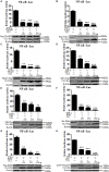
Inhibition of TNF-α–induced NF-κB activation by HSV-1 UL2. (A) HEK293T cells were transfected with promoter reporter plasmids NF-κB–Luc and pRL-TK, together with 500 ng of Flag empty vector or pUL2-Flag plasmid. Twenty-four hours posttransfection, cells were treated with or without 10 ng/mL of the recombinant human TNF-α and incubated for an additional 6 h, followed by cell lysed. Nuclear factor κB–driven luciferase activity was detected by DLR, as described in section “Materials and Methods.” (B) was carried out as (A); except that for an increase indicated amounts (100, 250, and 500 ng) of UL2-Flag expression plasmid were used. Cell lysates were divided into two aliquots; one aliquot was used for DLR detection, and the other was used for WB analysis to detect the protein expression of transfected plasmid. The expression of UL2 was analyzed by WB with anti-Flag mAb, and β-actin was used to verify equal loading of protein in each lane. Dual-luciferase reporter data were normalized for transfection efficiency through measuring firefly luciferase activity and Renilla luciferase activity, and values were shown as the ratio between the firefly and Renilla luciferase. Data were expressed as means ± SD from three independent experiments. ***P < 0.001.
FIGURE 2
FIGURE 2
Herpes simplex virus 1 UL2 inhibits NF-κB–driven cytokine expression. (A) HEK293T cells were cotransfected with Flag vector or diverse concentrations (100, 250, and 500 ng) of UL2-Flag expression plasmid along with reporter plasmids pXP2-pIL-8-Luc and pRL-TK. Twenty-four hours posttransfection, cells were treated with TNF-α (10 ng/mL) for 6 h, and luciferase activity was measured as described in Figure 1. (B) HEK293T cells were transfected with 1 μg of HA control vector or UL2-HA expression plasmid; 24 h posttransfection, cells were treated with TNF-α (10 ng/mL) for 6 h, and then RT-qPCR analysis was performed to analyze the relative expression level of IL-8 mRNA. Glyceraldehyde-3-phosphate dehydrogenase was used as the housekeeping gene. (C) was carried out as (B), except that HeLa cells were used for transfection. The expression of UL2 was analyzed by WB using anti-Flag mAb or anti-HA mAb, and β-actin was used to verify equal loading of protein in each lane. (D) HEK293T cells were mock-infected or infected with WT, UL2 Del, or UL2 Rev HSV-1 BAC GFP Luc virus at an MOI of 1 for 16 h. Flow cytometry analysis was then carried out to detect the GFP fluorescence. (E) HEK293T cells were infected with WT, UL2 Del, or UL2 Rev HSV-1 BAC GFP Luc virus at an MOI of 1. Sixteen hours postinfection, cells were treated with TNF-α (10 ng/mL) for 6 h. Then, RT-qPCR analysis was performed to detect the relative expression level of IL-8 mRNA. Data were expressed as means ± SD from three independent experiments. *P < 0.05, **P < 0.01, and ***P < 0.001.
FIGURE 3
FIGURE 3
Herpes simplex virus 1 UL2 targets at or downstream of the p65 level to suppress NF-κB pathway. HEK293T cells were cotransfected with reporter plasmids NF-κB–Luc, pRL-TK, and 100 ng of TRADD (A), TAK1 (B), TRAF2 (C), RIP1 (D), NIK (E), IKKα (F), IKKβ (G), or p65 (H) expression plasmid along with the indicated amounts (100, 250, and 500 ng) of UL2-Flag or UL2-HA expression plasmid, and then luciferase activity was analyzed as described in Figure 1. Cell lysates were analyzed by WB with corresponding tag-specific Abs to detect the expression of related plasmids, and β-actin was used to verify equal loading of protein in each lane. Data were expressed as means ± SD from three independent experiments. **P < 0.01 and ***P < 0.001.
FIGURE 4
FIGURE 4
Herpes simplex virus 1 UL2 interacts with endogenous p65 and p50. (A,C) HEK293T cells were cotransfected with pCMV-p65-Flag and pUL2-HA (A) or p50-Flag and pUL2-HA (C) expression plasmids. Twenty-four hours posttransfection, cells were harvested and lysed, and the samples were then subjected to Co-IP assays using anti-Flag mAb or non-specific mouse IgG. Western blots were probed with the indicated Abs. (B,D) HEK293T cells were cotransfected with plasmids as described for (A,C), respectively. Protein lysates were Co-IPed using anti-HA mAb, and WBs were analyzed with the indicated Abs. (E,F) HEK293T cells infected with WT HSV-1 at an MOI of 1 for 24 h were treated with 10 ng/mL of the TNF-α for an additional 6 h. Cells were then lysed, and the extracts were subjected to Co-IP using anti-p65 pAb (E), anti-p50 pAb (F), or control IgG. Samples were analyzed by WBs with the indicated Abs.
FIGURE 5
FIGURE 5
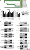
The region of aa9-17 is responsible for HSV-1 UL2 inhibition of NF-κB activity through interacting with p65 and p50. (A) Schematic representations of WT and truncated mutants of UL2 constructed in our laboratory. (B,C) HEK293T cells were cotransfected with NF-κB–Luc and pRL-TK reporter plasmids, along with 500 ng of EYFP empty vector or plasmid encoding EYFP-fused WT or truncated mutants of UL2. Twenty-four hours posttransfection, cells were treated with or without TNF-α (10 ng/mL) for 6 h, and luciferase activity was analyzed, as described in Figure 1. Data were expressed as means ± SD from three independent experiments. (D–N) HEK293T cells were cotransfected with pUL2(9-17)-EYFP and Flag vector (D), pUL2(9-17)-EYFP and pCMV-p65-Flag (E), pUL2(9-17)-EYFP and p50-Flag (F), pUL2(278-334)-EYFP and pCMV-p65-Flag (G), pUL2(278-334)-EYFP and p50-Flag (H), pUL2(9-17)del-EYFP and pCMV-p65-Flag (I), pUL2(9-17)del-EYFP and p50-Flag (J), pUL2(69-75)-EYFP and pCMV-p65-Flag (K), pUL2(69-75)-EYFP and p50-Flag (L), pUL2(9-17)(69-75)del-EYFP and pCMV-p65-Flag (M), or pUL2(9-17)(69-75)del-EYFP and p50-Flag (N) expression plasmids. Twenty-four hours posttransfection, cells were harvested and lysed, and the samples were then subjected to Co-IP assays using anti-Flag mAb, anti-EYFP or non-specific mouse IgG. Western blots were probed with the indicated Abs. **P < 0.01.
FIGURE 6
FIGURE 6
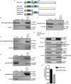
p65-IPT is associated with HSV-1 UL2. (A) Structural analysis diagrams of p65. RHD denotes Rel homology domain, and IPT denotes immunoglobulin-like plexin transcription factor. (B–F) HEK293T cells cotransfected with pUL2-HA and p65 truncated construct, including Pad-N-p65(1-290)-Flag (B), Pad-N-p65(291-551)-Flag (C), p65-IPT-EGFP (D), p65-RHD-Flag (E), or p65-IPT-del-Myc (F), were analyzed by Co-IP assays. (B,C,E) were Co-IPed with anti-Flag mAb; (D,F) were Co-IPed with anti-HA (D) and anti-Myc (F) mAbs, respectively. Western blots were probed with the indicated Abs. (G) HEK293T cells were cotransfected with expression plasmids UL2-Myc and p65-IPT-EGFP or EGFP vector. Twenty-four hours posttransfection, cell lysates were harvested and analyzed by Co-IP assays with anti-Myc mAb, and WBs were performed with the indicated Abs. Expression level of β-actin was served as loading control. (H) Densitometry of the UL2 and endogenous p65 protein interaction bands was normalized to β-actin. Data were expressed as means ± SD from three independent experiments. ***P < 0.001.
FIGURE 7
FIGURE 7
HSV-1 UL2 does not affect the dimerization of p65/p50. (A) HEK293T cells cotransfected with expression plasmids p65-EYFP, p50-Flag, and UL2-HA or HA vector construct for 24 h were harvested and analyzed by Co-IP assays using anti-Flag mAb, and WBs were performed using the indicated Abs. Expression level of β-actin was served as loading control. (B) Densitometry of the p65 and p50 interaction bands was normalized to β-actin. Data were expressed as means ± SD from three independent experiments. ns, not significant.
FIGURE 8
FIGURE 8
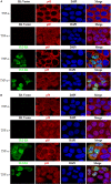
HSV-1 UL2 does not block the TNF-α–induced nuclear translocation of p65 or p50. HeLa cells were transfected with HA vector or UL2-HA expression plasmid. Twenty-four hours posttransfection, cells were treated with TNF-α (10 ng/mL) or mock-treated for 30 min. Then, cells were stained with anti-HA mAb and anti-p65 pAb (A) or anti-p50 pAb (B). Fluorescein isothiocyanate–conjugated donkey anti–mouse IgG (green) and Cy5-conjugated goat anti–rabbit IgG (red) were used as the secondary Abs. Cell nuclei were stained with DAPI (blue). All of the transfected cells were analyzed by a confocal microscope (Axio-Imager-LSM-800; Zeiss), and the photomicrographs were taken at a magnification of 400×. Each image represented a vast majority of the cells with similar subcellular distribution. Statistical analysis of the subcellular localization of p65 or p50 in the absence or presence of UL2 is shown in Table 2.
FIGURE 9
FIGURE 9
p65 phosphorylation at Ser536 is suppressed by HSV-1 UL2. (A,C) HEK293T cells transfected with either HA empty vector or UL2-HA expression plasmid were stimulated with TNF-α (20 ng/mL) for the indicated times (0, 30, and 60 min) according to previous studies (59, 77), and then equal amounts of cell lysates were analyzed by WBs with phospho-NF-κB–p65 (Ser536) Ab (A), phospho-NF-κB–p65 (Ser276) Ab (C) (top panel), or anti-p65 pAb (second panel). Protein levels of UL2 (third panel) and β-actin (bottom panel) in the same cell lysates were also determined. (B,D) Densitometry of phospho-NF-κB–p65 Ser536 (B) and Ser276 bands (D) from (A,C), respectively, were normalized to loading control β-actin. Data were expressed as means ± SD from three independent experiments. ns, not significant and ***P < 0.001.

Similar articles

Cited by

References

    1. Steiner I, Benninger F. Update on herpes virus infections of the nervous system. Curr Neurol Neurosci Rep. (2013) 13:414. 10.1007/s11910-013-0414-8 - DOI - PubMed
    1. Evans CM, Kudesia G, McKendrick M. Management of herpesvirus infections. Int J Antimicrob Agents. (2013) 42:119-128. 10.1016/j.ijantimicag.2013.04.023 - DOI - PubMed
    1. Bernstein DI, Bellamy AR, Hook EW, III, Levin MJ, Wald A, Ewell MG, et al. Epidemiology, clinical presentation, and antibody response to primary infection with herpes simplex virus type 1 and type 2 in young women. Clin Infect Dis. (2013) 56:344-351. 10.1093/cid/cis891 - DOI - PMC - PubMed
    1. Kwon MS, Carnt NA, Truong NR, Pattamatta U, White AJ, Samarawickrama C, et al. Dendritic cells in the cornea during Herpes simplex viral infection and inflammation. Surv Ophthalmol. (2017) 63:565-578. 10.1016/j.survophthal.2017.11.001 - DOI - PubMed
    1. Bogani F, Chua CN, Boehmer PE. Reconstitution of uracil DNA glycosylase-initiated base excision repair in herpes simplex virus-1. J Biol Chem. (2009) 284:16784-16790. 10.1074/jbc.M109.010413 - DOI - PMC - PubMed

Publication types

MeSH terms