. 2020 May 6:11:648.
doi: 10.3389/fimmu.2020.00648. eCollection 2020.

Interplay Between Keratinocytes and Fibroblasts: A Systematic Review Providing a New Angle for Understanding Skin Fibrotic Disorders

Affiliations

Interplay Between Keratinocytes and Fibroblasts: A Systematic Review Providing a New Angle for Understanding Skin Fibrotic Disorders

Barbara Russo et al. Front Immunol. .

Abstract

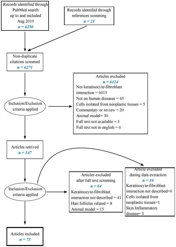
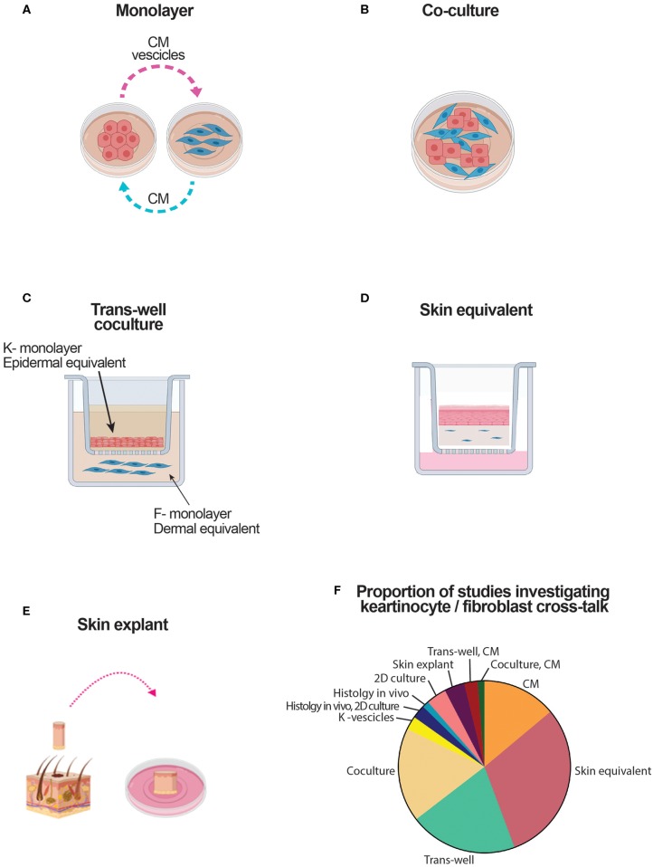
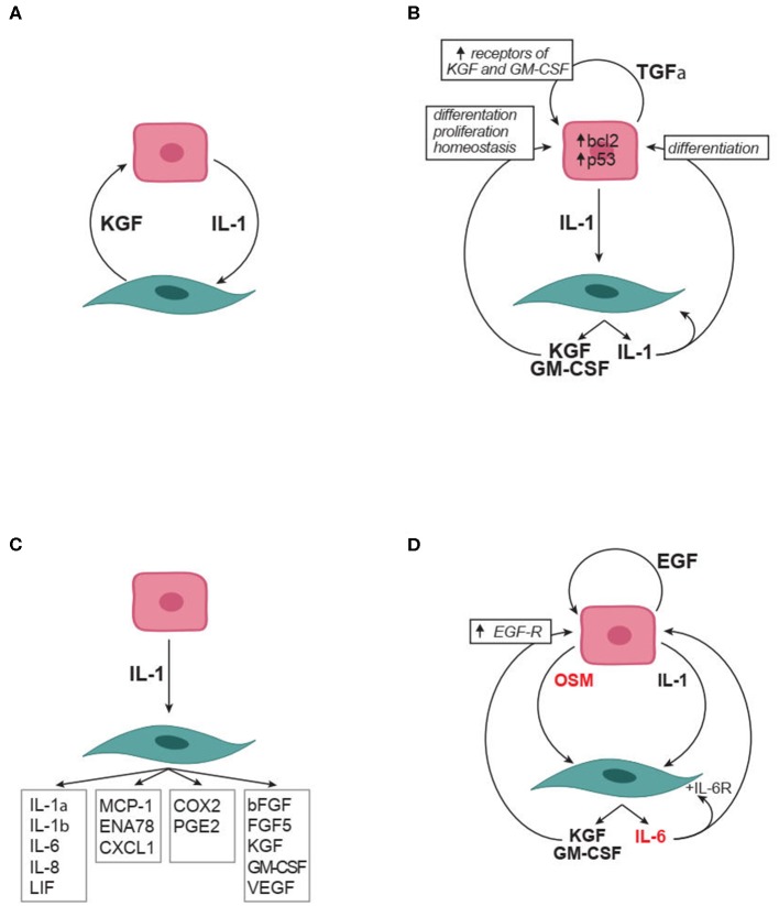
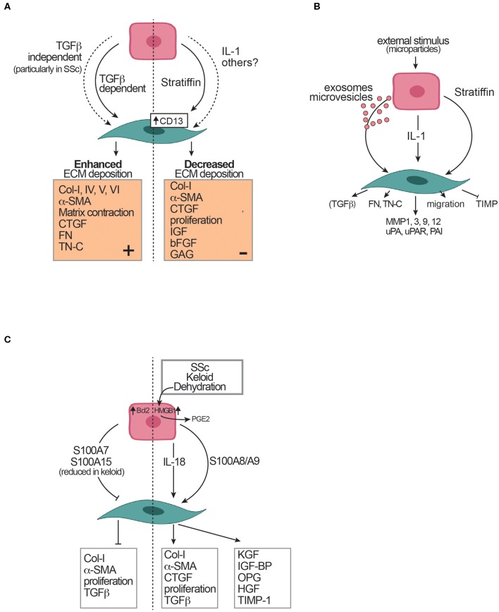
Background/Objective: Skin fibrosis is the result of aberrant processes leading to abnormal deposition of extracellular matrix (ECM) in the dermis. In healthy skin, keratinocytes participate to maintain skin homeostasis by actively crosstalking with fibroblasts. Within the wide spectrum of fibrotic skin disorders, relatively little attention has been devoted to the role of keratinocytes for their capacity to participate to skin fibrosis. This systematic review aims at summarizing the available knowledge on the reciprocal interplay of keratinocytes with fibroblasts and their soluble mediators in physiological states, mostly wound healing, and conditions associated with skin fibrosis. Methods: We performed a systematic literature search on PubMed to identify in vitro and ex vivo human studies investigating the keratinocyte characteristics and their interplay with fibroblasts in physiological conditions and within fibrotic skin disorders including hypertrophic scars, keloids, and systemic sclerosis. Studies were selected according to pre-specified eligibility criteria. Data on study methods, models, stimuli and outcomes were retrieved and summarized according to pre-specified criteria. Results: Among the 6,271 abstracts retrieved, 73 articles were included, of which 14 were specifically dealing with fibrotic skin pathologies. Fifty-six studies investigated how keratinocyte may affect fibroblast responses in terms of ECM-related genes or protein production, phenotype modification, and cytokine production. Most studies in both physiological conditions and fibrosis demonstrated that keratinocytes stimulate fibroblasts through the production of interleukin 1, inducing keratinocyte growth factor (KGF) and metalloproteinases in the fibroblasts. When the potential of keratinocytes to modulate collagen synthesis by healthy fibroblasts was explored, the results were controversial. Nevertheless, studies investigating keratinocytes from fibrotic skin, including keloids, hypertrophic scar, and scleroderma, suggested their potential involvement in enhancing ECM deposition. Twenty-three papers investigated keratinocyte proliferation differentiation and production of soluble mediators in response to interactions with fibroblasts. Most studies showed that fibroblasts modulate keratinocyte viability, proliferation, and differentiation. The production of KGF by fibroblast was identified as key for these functions. Conclusions: This review condenses evidence for the active interaction between keratinocytes and fibroblasts in maintaining skin homeostasis and the altered homeostatic interplay between keratinocytes and dermal fibroblasts in scleroderma and scleroderma-like disorders.

Keywords: cytokine; extracellular matrix; fibroblast; fibrosis; homeostasis; keratinocyte; systemic sclerosis.


Figures

Figure 1
Figure 1
Flow chart of the literature-searching strategy.
Figure 2
Figure 2
Schematic representation of culture systems used to assess the crosstalk between keratinocytes and fibroblasts. CM, conditioned medium; DE, dermal equivalent; EE, epidermal equivalent; F, fibroblast; K, keratinocyte. (A) Culture based on the use of medium conditioned by one type of cell cultured in monolayer to modulate the response of the other cell type. (B) Culture based on a mix of keratinocytes and fibroblasts. (C) Culture based on the use of transwells. Keratinocytes either in monolayer, either in dermal equivalents are in the top well. Fibroblasts, either in monolayer either in dermal equivalent are in the bottom well. Soluble mediators cross the semipermeable transwell membrane. (D) Skin equivalent generated at the air liquid interphase. (E) Organotypic full skin culture obtained by skin biopsy. (F) Proportion of the studies addressing keratinocyte-fibroblast crosstalk which results are reviewed here.
Figure 3
Figure 3
Soluble factors in the crosstalk between keratinocytes and fibroblasts. (A) IL-1 and KGF are soluble mediators robustly identified as involved in the crosstalk. (B) Autocrine and paracrine effects relevant to IL-1 and KGF role in the crosstalk. (C) The many effects of keratinocyte produced IL-1 on fibroblasts. (D) Synergistic effect of IL-1 with IL-6 family members (in red) on the crosstalk. Arrowheads indicate enhancement. bFGF, basic fibroblast growth factor; COX2, Cyclooxygenase 2; CXCL, chemokine containing the CXC motif; EGF, epidermal growth factor which comprises multiple mediators including transforming growth factor-α, amphiregulin, heparin binding-EGF, and epiregulin; ENA-78, Epithelial neutrophil-activating protein 78; FGF, fibroblast growth factor; GM-CSF, granulocyte-monocyte colony stimulating factor IL, interleukin; KGF, keratinocyte growth factor (also known as FGF7); LIF, leukemia inhibitory factor; MCP-1, monocyte chemotactic protein-1; OSM, oncostatin M;PGE2, Prostaglandin E2; TGF, Transforming growth factor; VEGF, vascular endothelial growth factor.
Figure 4
Figure 4
Effects of keratinocytes on fibroblasts and extracellular matrix (ECM). (A) Controversial effects of keratinocytes on ECM deposition. (B) Mediators of keratinocyte effects on fibroblasts. (C) Skin pathological conditions and their effects on the crosstalk between keratinocytes and fibroblasts. The dotted vertical line separates controversial evidence. Arrowheads indicate enhancement. Blunted heads indicate inhibition. αSMA, alpha-smooth muscle actin; Bcl2, B-cell lymphoma 2; bFGF, basic fibroblast growth factor; Col, collagen; CTGF, connective tissue growth factor; FN, fibronectin; HGF, hepatocyte growth factor; HMGB1, high mobility group box-1; IGF, insulin-like growth factor; IGF-BP, insulin-like growth factor binding protein; KGF, keratinocyte growth factor; MMP, metalloproteinase; OPG, osteoprotegerin; OSM, oncostatin M; PAI, plasminogen activator inhibitor; PDGF, platelet-derived growth factor; PGE2, Prostaglandin E2; S100A7, psoriasin; S100A8/A9, calprotectin; S100A15,koebnerisin; SSc, Systemic sclerosis; SSc-F, SSc fibroblasts; SSc-K, SSc keratinocytes; TGF, Transforming growth factor; TIMP, tissue inhibitor of MMP; TN-C, tenascin C; TNFα, Tumor necrosis factor α; uPA, urokinase-type plasminogen activator; uPAR, urokinase-type plasminogen activator receptor.
Figure 5
Figure 5
Effects of fibroblasts on keratinocytes. Arrowheads indicate enhancement. Blunted heads indicate inhibition. BM, basal membrane; HB-EGF, heparin binding EGF like growth factor; HGF, hepatocyte growth factor; IL, interleukin; KGF, keratinocyte growth factor; PDGF, platelet derived growth factor; PGE2, prostaglandin E2; PPAR, peroxisome proliferator-activated receptor; TGF, transforming growth factor.

Similar articles

Cited by

References

    1. Rockey DC, Bell PD, Hill JA. Fibrosis–a common pathway to organ injury and failure. N Engl J Med. (2015) 372:1138–49. 10.1056/NEJMra1300575 - DOI - PubMed
    1. Distler JHW, Gyorfi AH, Ramanujam M, Whitfield ML, Konigshoff M, Lafyatis R. Shared and distinct mechanisms of fibrosis. Nat Rev Rheumatol. (2019) 15:705–30. 10.1038/s41584-019-0322-7 - DOI - PubMed
    1. Duffield JS, Lupher M, Thannickal VJ, Wynn TA. Host responses in tissue repair and fibrosis. Annu Rev Pathol. (2013) 8:241–76. 10.1146/annurev-pathol-020712-163930 - DOI - PMC - PubMed
    1. Boin F, Chizzolini C. Inflammation and immunity. In: Varga J, Denton C, Wigley F, Allanore Y, Kuwana M, editors. Scleroderma: From Pathogenesis to Comprehensive Management. New York, NY: Springer; (2017). p. 161–96.
    1. Lodyga M, Hinz B. TGF-β–a truly transforming growth factor in fibrosis and immunity. Semin Cell Dev Biol. (2020) 101:123–39. 10.1016/j.semcdb.2019.12.010 - DOI - PubMed

Publication types