

A report of two challenging cases of bone infection: Mycobacterium tuberculosis. How to manage?
- PMID: 32477575
- PMCID: PMC7243712
- DOI: 10.1093/omcr/omaa025
A report of two challenging cases of bone infection: Mycobacterium tuberculosis. How to manage?
Abstract
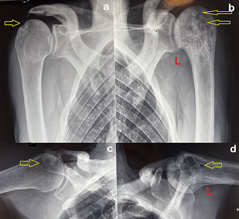
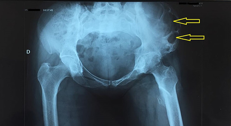
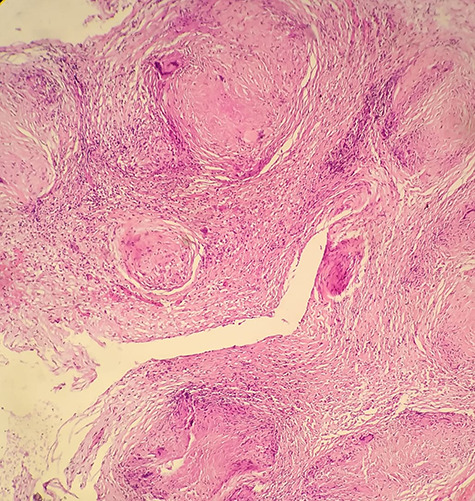
The incidence of bone tuberculosis is less than 5% of all tuberculosis cases. Furthermore, multifocal bone tuberculosis is uncommon, which rarely occurs without primary foci. It is difficult to diagnose, particularly if it is localized in both humeral heads. On the other hand, the isolated iliac bone tuberculosis is exceptional; it constitutes, also, a challenging diagnosis, which requires a high index of clinical suspicion and advanced investigations. Herein, we first report a case of multifocal tuberculosis of both humeral heads with no primary foci, and we secondarily report a case of isolated iliac bone tuberculosis. At last, however, the histological exam and polymerase chain reaction for the Mycobacterium tuberculosis complex are not always positives; they are mandatory as tests to ascertain the diagnosis.
Keywords: anti-bacillary; bone tuberculosis; polymerase chain reaction.
© The Author(s) 2020. Published by Oxford University Press.
Figures


Similar articles
-
A Case of Generalized, Superinfected Dermatitis and Inguinal Mycobacterium Lymphadenitis - TB or not TB?Acta Dermatovenerol Croat. 2018 Oct;26(3):270-272. Acta Dermatovenerol Croat. 2018. PMID: 30390733
-
Multifocal tuberculosis simulating a cancer-a case report.BMC Infect Dis. 2020 Jul 10;20(1):495. doi: 10.1186/s12879-020-05209-x. BMC Infect Dis. 2020. PMID: 32650727 Free PMC article.
-
[Multifocal bone tuberculosis: apropos of a case].Rev Chir Orthop Reparatrice Appar Mot. 1995;81(6):553-6. Rev Chir Orthop Reparatrice Appar Mot. 1995. PMID: 8560027 French.
-
Perianal Tuberculosis: Lessons Learned in 57 Patients From 743 Samples of Histopathology and Polymerase Chain Reaction and a Systematic Review of Literature.Dis Colon Rectum. 2019 Nov;62(11):1390-1400. doi: 10.1097/DCR.0000000000001493. Dis Colon Rectum. 2019. PMID: 31596764
-
[Development of antituberculous drugs: current status and future prospects].Kekkaku. 2006 Dec;81(12):753-74. Kekkaku. 2006. PMID: 17240921 Review. Japanese.
Cited by
-
Tuberculosis of the Iliac Bone and Acetabulum With Iliopsoas, Obturator Internus, and Obturator Externus Abscesses in an Immunocompetent Indian Female: An Extremely Rare Case.Cureus. 2024 Mar 7;16(3):e55727. doi: 10.7759/cureus.55727. eCollection 2024 Mar. Cureus. 2024. PMID: 38586629 Free PMC article.
-
Epidemiological and osteoarticular involvement sites' characteristics of multiple osteoarticular tuberculosis: a scoping review.Epidemiol Infect. 2025 Jan 21;153:e26. doi: 10.1017/S095026882400150X. Epidemiol Infect. 2025. PMID: 39834064 Free PMC article.
-
Iliac Bone Tuberculosis Presenting as Left Thigh Swelling in an Indian Female Patient: A Rare Case.Cureus. 2022 Aug 23;14(8):e28297. doi: 10.7759/cureus.28297. eCollection 2022 Aug. Cureus. 2022. PMID: 36158372 Free PMC article.
-
Pediatric Osteoarticular Tuberculosis as a Diagnostic Dilemma and a Review of Literature.Cureus. 2022 Mar 11;14(3):e23053. doi: 10.7759/cureus.23053. eCollection 2022 Mar. Cureus. 2022. PMID: 35308187 Free PMC article.
References
-
- Stingo FE, Rodriguez-Fontan F, Burger-Van der Walt E, Arce J, Garcia SN, Munafo RM. Isolated iliac crest tuberculosis: a case report. JBJS Case Connect 2018;8:e31. - PubMed
-
- Yilmaz MH, Kantarci F, Mihmanli I, Kanberoglu K. Multifocal skeletal tuberculosis. South Med J 2004;97:785–7. - PubMed
-
- Davidson PT, Horowitz I. Skeletal tuberculosis: a review with patient presentations and discussion. Am J Med 1970;48:77–84. - PubMed
LinkOut - more resources
Full Text Sources
