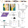
Effect of Acellular Amnion With Increased TGF-β and bFGF Levels on the Biological Behavior of Tenocytes
- PMID: 32478059
- PMCID: PMC7240037
- DOI: 10.3389/fbioe.2020.00446
Effect of Acellular Amnion With Increased TGF-β and bFGF Levels on the Biological Behavior of Tenocytes
Abstract
The human amniotic membrane has been a subject for clinical and basic research for nearly 100 years, but weak rejection has been reported. The purpose of this research is to remove the cellular components of the amnion for eliminating its immune-inducing activity to the utmost extent. The amniotic membrane treated by acid removed the epithelial cell, fibroblast, and sponge layers and retained only the basal and dense layers. In vitro, biological effects of the new material on tenocytes were evaluated. The levels of transforming growth factor (TGF-β1), fibroblast growth factor (bFGF) proteins were measured. In vivo, the tendon injury model of chickens was constructed to observe effects on tendon adhesion and healing. The acellular amniotic membrane effectively removed the cell components of the amnion while retaining the fibrous reticular structure. Abundant collagen fibers enhanced the tensile strength of amnion, and a 3D porous structure provided enough 3D space structure for tenocyte growth. In vitro, acellular amnion resulted in the fast proliferation trend for tenocytes with relatively static properties by releasing TGF-β1 and bFGF. In vivo, the experiment revealed the mechanism of acellular amnion in promoting endogenous healing and barrier exogenous healing by evaluating tendon adhesion, biomechanical testing, and labeling fibroblasts/tendon cells and monocytes/macrophages with vimentin and CD68. The acellular amnion promotes endogenous healing and barrier exogenous healing by releasing the growth factors such as TGF-β1 and bFGF, thereby providing a new direction for the prevention and treatment of tendon adhesion.
Keywords: amnion; collagen; growth factor; tendon adhesions; tenocytes.
Copyright © 2020 Sang, Liu, Kong, Qian and Liu.
Figures


Similar articles
-
Regulation of ERK1/2 and SMAD2/3 Pathways by Using Multi-Layered Electrospun PCL-Amnion Nanofibrous Membranes for the Prevention of Post-Surgical Tendon Adhesion.Int J Nanomedicine. 2020 Feb 11;15:927-942. doi: 10.2147/IJN.S231538. eCollection 2020. Int J Nanomedicine. 2020. PMID: 32103947 Free PMC article.
-
Evaluation of two distinct placental-derived membranes and their effect on tenocyte responses in vitro.J Tissue Eng Regen Med. 2019 Aug;13(8):1316-1330. doi: 10.1002/term.2876. Epub 2019 Jun 13. J Tissue Eng Regen Med. 2019. PMID: 31062484 Free PMC article.
-
Tendon healing in vitro: bFGF gene transfer to tenocytes by adeno-associated viral vectors promotes expression of collagen genes.J Hand Surg Am. 2005 Nov;30(6):1255-61. doi: 10.1016/j.jhsa.2005.06.001. J Hand Surg Am. 2005. PMID: 16344185
-
The Functions and Mechanisms of Basic Fibroblast Growth Factor in Tendon Repair.Front Physiol. 2022 Jun 13;13:852795. doi: 10.3389/fphys.2022.852795. eCollection 2022. Front Physiol. 2022. PMID: 35770188 Free PMC article. Review.
-
The roles of growth factors in tendon and ligament healing.Sports Med. 2003;33(5):381-94. doi: 10.2165/00007256-200333050-00004. Sports Med. 2003. PMID: 12696985 Review.
Cited by
-
Interposition of acellular amniotic membrane at the tendon to bone interface would be better for healing than overlaying above the tendon to bone junction in the repair of rotator cuff injury.Chin J Traumatol. 2025 May;28(3):187-192. doi: 10.1016/j.cjtee.2024.04.001. Epub 2024 Apr 20. Chin J Traumatol. 2025. PMID: 38688817 Free PMC article.
-
Dietary Aflatoxin B1 attenuates immune function of immune organs in grass carp (Ctenopharyngodon idella) by modulating NF-κB and the TOR signaling pathway.Front Immunol. 2022 Oct 18;13:1027064. doi: 10.3389/fimmu.2022.1027064. eCollection 2022. Front Immunol. 2022. PMID: 36330527 Free PMC article.
-
Human Acellular Amniotic Membrane as Skin Substitute and Biological Scaffold: A Review of Its Preparation, Preclinical Research, and Clinical Application.Pharmaceutics. 2023 Aug 30;15(9):2249. doi: 10.3390/pharmaceutics15092249. Pharmaceutics. 2023. PMID: 37765218 Free PMC article. Review.
-
Historical evolution, hotspots, and trends in tendon tissue engineering: A bibliometric analysis.Regen Ther. 2025 May 6;29:600-612. doi: 10.1016/j.reth.2025.04.009. eCollection 2025 Jun. Regen Ther. 2025. PMID: 40487919 Free PMC article.
-
Viewing Decellularized Amniotic Membrane Through the Lens of Coupled Scaffolding and Drug Delivery Systems in Regenerative Medicine.J Tissue Eng Regen Med. 2025 Jul 23;2025:8818058. doi: 10.1155/term/8818058. eCollection 2025. J Tissue Eng Regen Med. 2025. PMID: 40741540 Free PMC article. Review.
References
-
- Butler D. L., Juncosa-Melvin N., Boivin G. P., Galloway M. T., Shearn J. T., Gooch C., et al. (2008). Functional tissue engineering for tendon repair: a multidisciplinary strategy using mesenchymal stem cells, bioscaffolds, and mechanical stimulation. J. Orthop. Res. 26 1–9. 10.1002/jor.20456 - DOI - PubMed
-
- Chen Y. J., Chung M. C., Jane Yao C. C., Huang C. H., Chang H. H., Jeng J. H., et al. (2012). The effects of acellular amniotic membrane matrix on osteogenic differentiation and erk1/2 signaling in human dental apical papilla cells. Biomaterials 33 455–463. 10.1016/j.biomaterials.2011.09.065 - DOI - PubMed
LinkOut - more resources
Full Text Sources

