

Structural Basis for Targeting T:T Mismatch with Triaminotriazine-Acridine Conjugate Induces a U-Shaped Head-to-Head Four-Way Junction in CTG Repeat DNA
- PMID: 32478511
- PMCID: PMC7837310
- DOI: 10.1021/jacs.0c03591
Structural Basis for Targeting T:T Mismatch with Triaminotriazine-Acridine Conjugate Induces a U-Shaped Head-to-Head Four-Way Junction in CTG Repeat DNA
Abstract
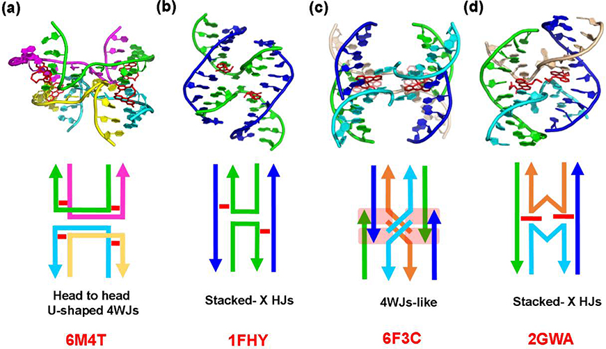
The potent DNA-binding compound triaminotriazine-acridine conjugate (Z1) functions by targeting T:T mismatches in CTG trinucleotide repeats that are responsible for causing neurological diseases such as myotonic dystrophy type 1, but its binding mechanism remains unclear. We solved a crystal structure of Z1 in a complex with DNA containing three consecutive CTG repeats with three T:T mismatches. Crystallographic studies revealed that direct intercalation of two Z1 molecules at both ends of the CTG repeat induces thymine base flipping and DNA backbone deformation to form a four-way junction. The core of the complex unexpectedly adopts a U-shaped head-to-head topology to form a crossover of each chain at the junction site. The crossover junction is held together by two stacked G:C pairs at the central core that rotate with respect to each other in an X-shape to form two nonplanar minor-groove-aligned G·C·G·C tetrads. Two stacked G:C pairs on both sides of the center core are involved in the formation of pseudo-continuous duplex DNA. Four metal-mediated base pairs are observed between the N7 atoms of G and CoII, an interaction that strongly preserves the central junction site. Beyond revealing a new type of ligand-induced, four-way junction, these observations enhance our understanding of the specific supramolecular chemistry of Z1 that is essential for the formation of a noncanonical DNA superstructure. The structural features described here serve as a foundation for the design of new sequence-specific ligands targeting mismatches in the repeat-associated structures.
Figures


Similar articles
-
Selective alkylation of T-T mismatched DNA using vinyldiaminotriazine-acridine conjugate.Nucleic Acids Res. 2018 Feb 16;46(3):1059-1068. doi: 10.1093/nar/gkx1278. Nucleic Acids Res. 2018. PMID: 29309639 Free PMC article.
-
Binding of a macrocyclic bisacridine and ametantrone to CGTACG involves similar unusual intercalation platforms.Biochemistry. 2000 Sep 12;39(36):10950-7. Biochemistry. 2000. PMID: 10998231
-
A simple ligand that selectively targets CUG trinucleotide repeats and inhibits MBNL protein binding.Proc Natl Acad Sci U S A. 2009 Sep 22;106(38):16068-73. doi: 10.1073/pnas.0901824106. Epub 2009 Sep 8. Proc Natl Acad Sci U S A. 2009. PMID: 19805260 Free PMC article.
-
Unusual DNA duplex and hairpin motifs.Nucleic Acids Res. 2003 May 15;31(10):2461-74. doi: 10.1093/nar/gkg367. Nucleic Acids Res. 2003. PMID: 12736295 Free PMC article. Review.
-
CTG repeats associated with human genetic disease are inherently flexible.J Mol Biol. 1998 Jan 23;275(3):405-11. doi: 10.1006/jmbi.1997.1502. J Mol Biol. 1998. PMID: 9466918 Review.
Cited by
-
A Novel Minor Groove Binder as a Potential Therapeutic Agent for Myotonic Dystrophy Type 1.ChemMedChem. 2021 Sep 6;16(17):2638-2644. doi: 10.1002/cmdc.202100243. Epub 2021 Jun 10. ChemMedChem. 2021. PMID: 34114350 Free PMC article.
-
The role of water in mediating DNA structures with epigenetic modifications, higher-order conformations and drug-DNA interactions.RSC Chem Biol. 2025 Mar 14;6(5):699-720. doi: 10.1039/d4cb00308j. eCollection 2025 May 8. RSC Chem Biol. 2025. PMID: 40171245 Free PMC article. Review.
-
Staggered intercalation of DNA duplexes with base-pair modulation by two distinct drug molecules induces asymmetric backbone twisting and structure polymorphism.Nucleic Acids Res. 2022 Aug 26;50(15):8867-8881. doi: 10.1093/nar/gkac629. Nucleic Acids Res. 2022. PMID: 35871296 Free PMC article.
-
Recognition of mismatched sites in double-stranded DNA by a pair of partially noncomplementary peptide nucleic acids.Chem Lett. 2024 Dec 10;53(12):upae234. doi: 10.1093/chemle/upae234. eCollection 2024 Dec. Chem Lett. 2024. PMID: 39677325 Free PMC article.
-
Targeting the ALS/FTD-associated A-DNA kink with anthracene-based metal complex causes DNA backbone straightening and groove contraction.Nucleic Acids Res. 2021 Sep 20;49(16):9526-9538. doi: 10.1093/nar/gkab227. Nucleic Acids Res. 2021. PMID: 33836081 Free PMC article.
References
-
- Li G-M Mechanisms and functions of DNA mismatch repair. Cell Res. 2008, 18, 85–98. - PubMed
-
- Li GM Mechanisms and functions of DNA mismatch repair. Cell Res. 2008, 18, 85–98. - PubMed
-
- Tseng W-H; Chang C.-k.; Wu P-C; Hu N-J; Lee G-H; Tzeng C-C; Neidle S; Hou M-H Induced-Fit Recognition of CCG Trinucleotide Repeats by a Nickel–Chromomycin Complex Resulting in Large-Scale DNA Deformation. Angew. Chem. Int. Ed. 2017, 56, 8761–8765. - PubMed
Publication types
MeSH terms
Substances
Grants and funding
LinkOut - more resources
Full Text Sources
