

Prevalence of chondrocalcinosis in the temporomandibular joint in patients with chondrocalcinosis of the knee or wrist
- PMID: 32479114
- PMCID: PMC7549530
- DOI: 10.1259/dmfr.20190450
Prevalence of chondrocalcinosis in the temporomandibular joint in patients with chondrocalcinosis of the knee or wrist
Abstract
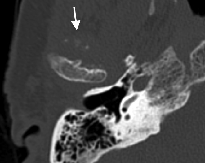
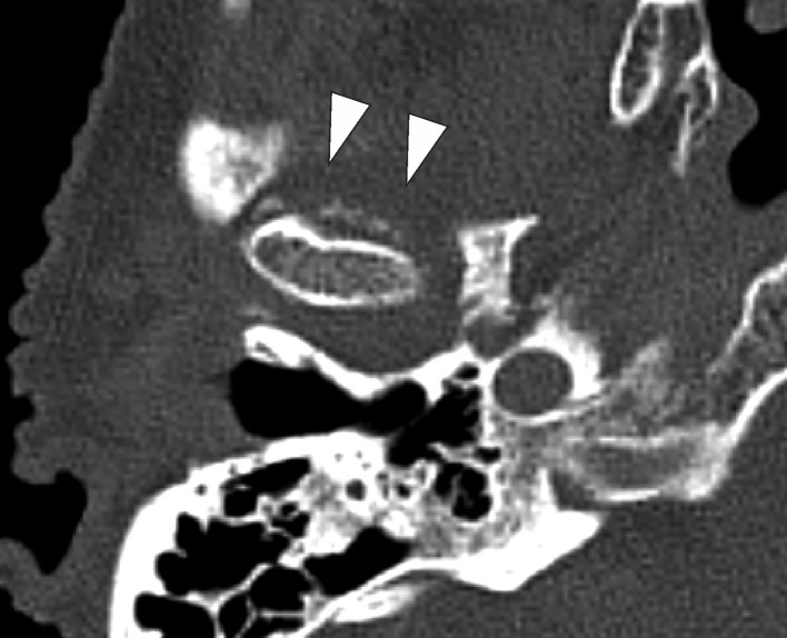
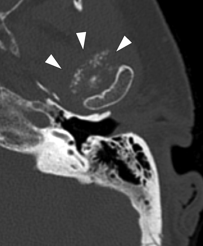
Objective: The aim of this study was to investigate the prevalence of TMJ chondrocalcinosis on head CT scans in patients with chondrocalcinosis of the knee or wrist.
Methods and materials: 227 patients with radiological evidence of calcifications on knee or wrist radiographs had a head CT scan obtained for unrelated purposes. CT scans were retrospectively reviewed for the presence of temporomandibular crystal deposition. Prevalence, bilaterality, age and gender distribution were determined.
Results: 41 of 227 (18%) of patients had TMJ chondrocalcinosis. TMJ chondrocalcinosis was more common in females (17%) than males (1%). It was more commonly unilateral (68%) than bilateral (32%).
Conclusion: In patients with peripheral calcific disease, the TMJ is more commonly involved than previously reported and this is more common in females compared to males.
Keywords: CPPD; CT; Chondrocalcinosis; Joint calcification; Temporomandibular Joint.
Figures
Similar articles
-
Pseudogout of the temporomandibular joint: a case report with systematic literature review.J Oral Facial Pain Headache. 2025 Mar;39(1):49-69. doi: 10.22514/jofph.2025.004. Epub 2025 Mar 12. J Oral Facial Pain Headache. 2025. PMID: 40129423 Free PMC article.
-
Cartilage icing and chondrocalcinosis on knee radiographs in the differentiation between gout and calcium pyrophosphate deposition.PLoS One. 2020 Apr 16;15(4):e0231508. doi: 10.1371/journal.pone.0231508. eCollection 2020. PLoS One. 2020. PMID: 32298308 Free PMC article.
-
Does chondrocalcinosis affect Knee Society scores and range of motion after TKA?Clin Orthop Relat Res. 2014 May;472(5):1512-7. doi: 10.1007/s11999-013-3447-z. Epub 2014 Jan 3. Clin Orthop Relat Res. 2014. PMID: 24385044 Free PMC article.
-
Chondrocalcinosis is common in the absence of knee involvement.Arthritis Res Ther. 2012 Oct 4;14(5):R205. doi: 10.1186/ar4043. Arthritis Res Ther. 2012. PMID: 23036436 Free PMC article.
-
Imaging of chondrocalcinosis: calcium pyrophosphate dihydrate (CPPD) crystal deposition disease -- imaging of common sites of involvement.Clin Exp Rheumatol. 2012 Jan-Feb;30(1):118-25. Epub 2012 Mar 7. Clin Exp Rheumatol. 2012. PMID: 22325558 Review.
Cited by
-
Pseudogout of the temporomandibular joint: a case report with systematic literature review.J Oral Facial Pain Headache. 2025 Mar;39(1):49-69. doi: 10.22514/jofph.2025.004. Epub 2025 Mar 12. J Oral Facial Pain Headache. 2025. PMID: 40129423 Free PMC article.
References
-
- Textbook of Rheumatology. 9th. 2013. Kelley’s.
-
- Cassidy Petty: Textbook of Pediatric Rheumatology. 4th ed; 2001.
-
- Hamblen Simpson: Adams’s Outline of Orthopaedics. 14th ed; 2010.
-
- Abhishek A, Doherty M. Update on calcium pyrophosphate deposition. Clin Exp Rheumatol 2016; (34(Suppl 98): 32–8. Epub 2016 Jul 22Jul-Aug;34. - PubMed
