

COVID-19-Associated Encephalitis Mimicking Glial Tumor
- PMID: 32479911
- PMCID: PMC7256557
- DOI: 10.1016/j.wneu.2020.05.194
COVID-19-Associated Encephalitis Mimicking Glial Tumor
Abstract
Background: Reports on neurologic manifestations of coronavirus disease 2019 (COVID-19) have attracted broad attention. We present an unusual case of COVID-19-associated encephalitis mimicking a glial tumor.
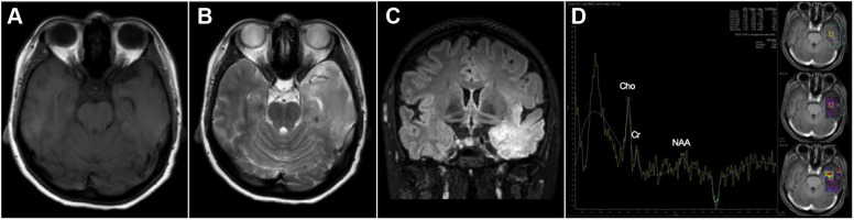
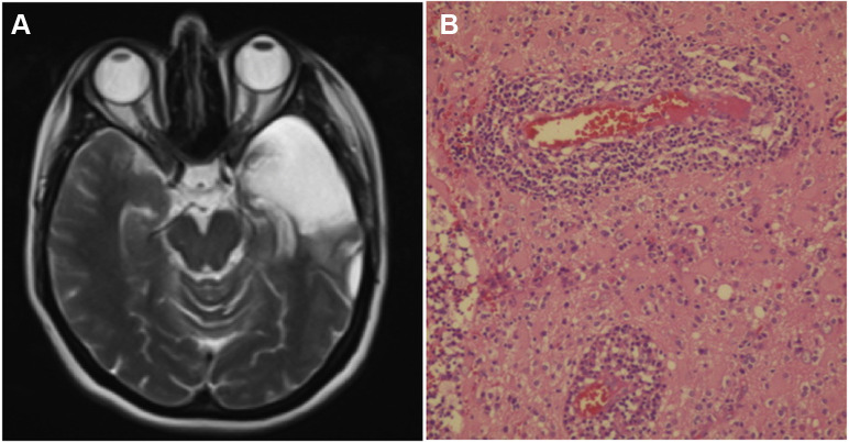
Case description: A 35-year-old woman presented with headache and seizures. T2 fluid-attenuated inverse recovery imaging showed hyperintensities in the left temporal lobe. Magnetic resonance spectroscopy showed an elevated choline peak. Imaging findings were suggestive of high-grade glioma. Antiepileptic medication failed to achieve seizure control. A left anterior temporal lobectomy was performed. The patient had no postoperative deficits, and her symptoms completely improved. Histologic examination revealed encephalitis. Postoperatively, our patient tested positive for COVID-19.
Conclusions: Our case raises awareness of neurologic manifestations of the disease and their potential to mimic glial tumors. For prompt diagnosis and prevention of transmission, clinicians should consider COVID-19 in patients with similar presentation.
Keywords: COVID-19; Coronavirus; Encephalitis; SARS-CoV-2; Spectroscopy.
Copyright © 2020 Elsevier Inc. All rights reserved.
Figures
Similar articles
-
Neurological manifestations of COVID-19: available evidences and a new paradigm.J Neurovirol. 2020 Oct;26(5):619-630. doi: 10.1007/s13365-020-00895-4. Epub 2020 Aug 24. J Neurovirol. 2020. PMID: 32839951 Free PMC article. Review.
-
[Encephalitis associated with COVID-19 in a 13-year-old girl: A case report].Medwave. 2020 Aug 3;20(7):e7984. doi: 10.5867/medwave.2020.07.7984. Medwave. 2020. PMID: 32804920 Spanish.
-
SARS-CoV-2-Associated Acute Hemorrhagic, Necrotizing Encephalitis (AHNE) Presenting with Cognitive Impairment in a 44-Year-Old Woman without Comorbidities: A Case Report.Am J Case Rep. 2020 Aug 16;21:e925641. doi: 10.12659/AJCR.925641. Am J Case Rep. 2020. PMID: 32799213 Free PMC article.
-
First motor seizure as presenting symptom of SARS-CoV-2 infection.Neurol Sci. 2020 Jul;41(7):1651-1653. doi: 10.1007/s10072-020-04460-z. Epub 2020 May 16. Neurol Sci. 2020. PMID: 32417987 Free PMC article. No abstract available.
-
Neurological complications of coronavirus and COVID-19.Rev Neurol. 2020 May 1;70(9):311-322. doi: 10.33588/rn.7009.2020179. Rev Neurol. 2020. PMID: 32329044 Review. English, Spanish.
Cited by
-
New-Onset Seizure With Possible Limbic Encephalitis in a Patient With COVID-19 Infection: A Case Report and Review.J Investig Med High Impact Case Rep. 2021 Jan-Dec;9:2324709620986302. doi: 10.1177/2324709620986302. J Investig Med High Impact Case Rep. 2021. PMID: 33648382 Free PMC article.
-
Acute and Post-Acute Neurological Complications of COVID-19.Neurol Int. 2021 Mar 9;13(1):102-119. doi: 10.3390/neurolint13010010. Neurol Int. 2021. PMID: 33803475 Free PMC article. Review.
-
Neurological manifestations and neuroimaging findings in patients with SARS-CoV2-a systematic review.Egypt J Neurol Psychiatr Neurosurg. 2021;57(1):68. doi: 10.1186/s41983-021-00322-3. Epub 2021 Jun 2. Egypt J Neurol Psychiatr Neurosurg. 2021. PMID: 34093004 Free PMC article. Review.
-
Neurological Complications of COVID-19 in the Elderly.Neurosci Behav Physiol. 2022;52(5):625-634. doi: 10.1007/s11055-022-01287-3. Epub 2022 Sep 13. Neurosci Behav Physiol. 2022. PMID: 36119647 Free PMC article.
-
Effects of COVID-19 on the Nervous System.Cell. 2020 Oct 1;183(1):16-27.e1. doi: 10.1016/j.cell.2020.08.028. Epub 2020 Aug 19. Cell. 2020. PMID: 32882182 Free PMC article. Review.
References
-
- Poyiadji N., Shahin G., Noujaim D., Stone M., Patel S., Griffith B. COVID-19–associated acute hemorrhagic necrotizing encephalopathy: CT and MRI features [e-pub ahead of print] https://doi.org/10.1148/radiol.2020201187 Radiology. - DOI - PMC - PubMed
-
- Tsai L.-K., Hsieh S.-T., Chang Y.-C. Neurological manifestations in severe acute respiratory syndrome. Acta Neurol Taiwanica. 2005;14:113–119. - PubMed
-
- Wu Y., Xu X., Chen Z. Nervous system involvement after infection with COVID-19 and other coronaviruses [e-pub ahead of print]. Brain Behav Immun. https://doi.org/10.1016/j.bbi.2020.03.031 Available at: - DOI - PMC - PubMed
Publication types
MeSH terms
LinkOut - more resources
Full Text Sources
Medical
Miscellaneous
