. 2020 Aug 15:401:115078.
doi: 10.1016/j.taap.2020.115078. Epub 2020 May 29.

A type IV collagenase inhibitor, N-hydroxy-3-phenyl-2-(4-phenylbenzenesulfonamido) propanamide (BiPS), suppresses skin injury induced by sulfur mustard

Affiliations

A type IV collagenase inhibitor, N-hydroxy-3-phenyl-2-(4-phenylbenzenesulfonamido) propanamide (BiPS), suppresses skin injury induced by sulfur mustard

Yoke-Chen Chang et al. Toxicol Appl Pharmacol. .

Abstract

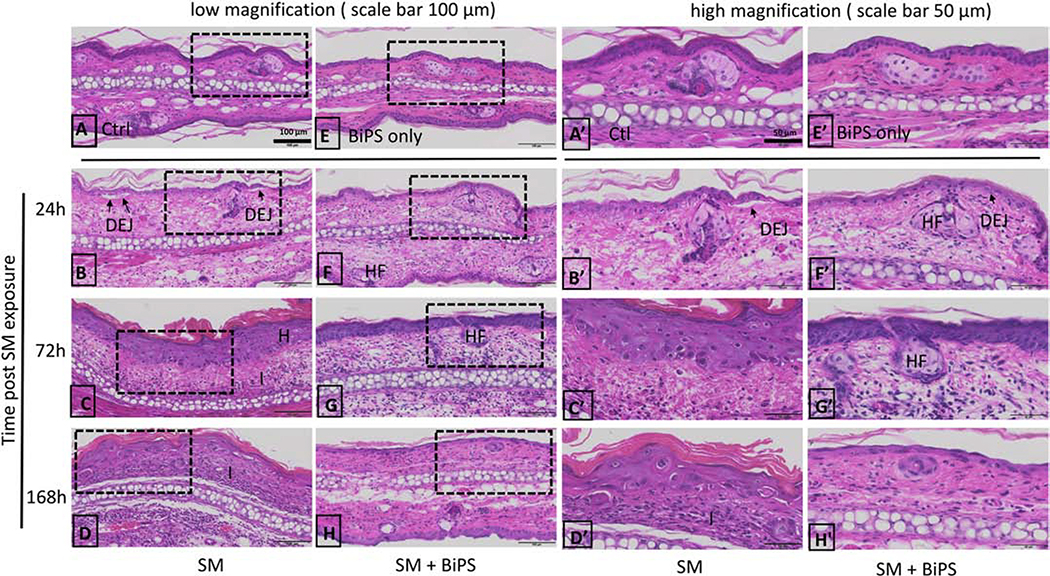
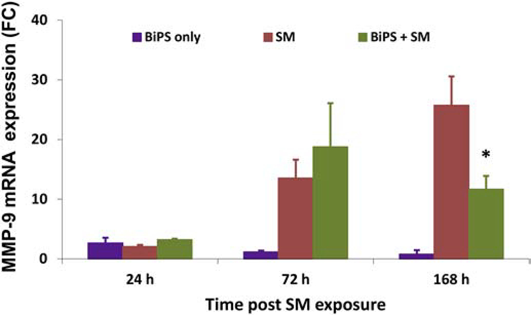
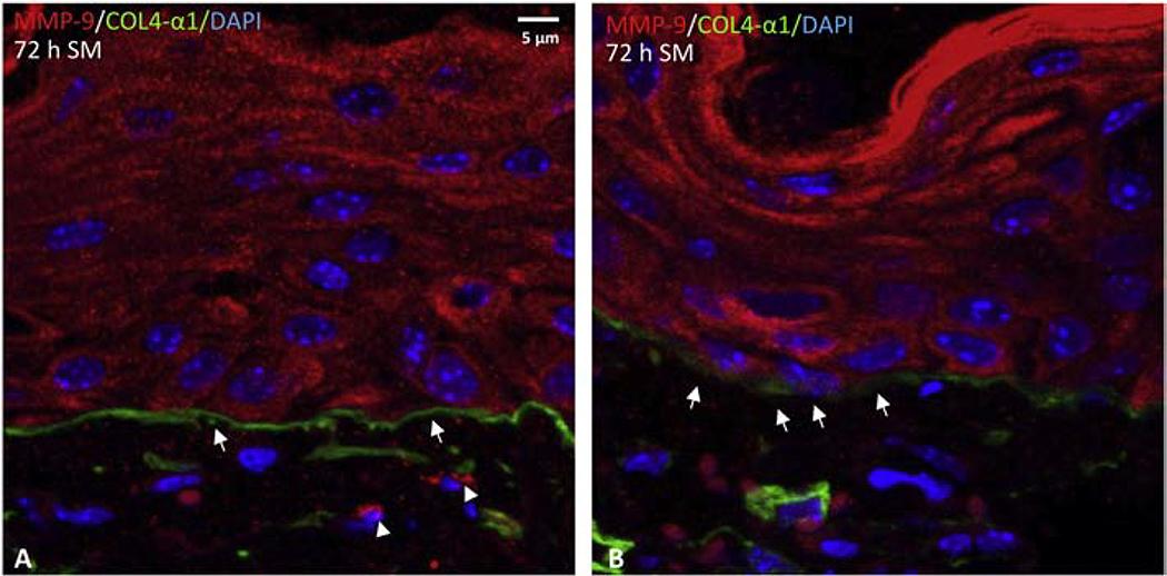
Sulfur mustard (SM) is a highly toxic blistering agent thought to mediate its action, in part, by activating matrix metalloproteinases (MMPs) in the skin and disrupting components of the basement membrane zone (BMZ). Type IV collagenases (MMP-9) degrade type IV collagen in the skin, a major component of the BMZ at the dermal-epidermal junction. In the present studies, a type IV collagenase inhibitor, N-hydroxy-3-phenyl-2-(4-phenylbenzenesulfonamido) propanamide (BiPS), was tested for its ability to protect the skin against injury induced by SM in the mouse ear vesicant model. SM induced inflammation, epidermal hyperplasia and microblistering at the dermal/epidermal junction of mouse ears 24-168 h post-exposure. This was associated with upregulation of MMP-9 mRNA and protein in the skin. Dual immunofluorescence labeling showed increases in MMP-9 in the epidermis and in the adjacent dermal matrix of the SM injured skin, as well as breakdown of type IV collagen in the basement membrane. Pretreatment of the skin with BiPS reduced signs of SM-induced cutaneous toxicity; expression of MMP-9 mRNA and protein was also downregulated in the skin by BiPS. Following BiPS pretreatment, type IV collagen appeared intact and was similar to control skin. These results demonstrate that inhibiting type IV collagenases in the skin improves basement membrane integrity after exposure to SM. BiPS may hold promise as a potential protective agent to mitigate SM induced skin injury.

Keywords: Basement Membrane; MMP-9; Matrix Metalloproteinase Inhibitor; Sulfur Mustard; Type IV Collagen.


Conflict of interest statement

Declaration of Competing Interest The authors declare that there are no conflicts of interest.

Figures

Fig. 1.
Fig. 1.. Chemical structure of BiPS.
BiPS [(2R)-[(4-biphenylylsulfonyl)amino]-N-hydroxy-3-phenylpropionamide] is an MMP-9 inhibitor.
Fig. 2.
Fig. 2.. Structural alterations in mouse ear skin following SM exposure.
H&E stained histological sections of control mouse ear skin and SM-treated mouse ear skin. Images shown in Panels A-H are at low magnification (scale bar = 100 μm). Images shown in Panels A’-H’ are high magnification (scale bar = 50 μm); Panels A’-H’ show images from the dashed boxed area of images panels A-H. Tissues were collected 24 h, 72 h and 168 h post-SM exposure. Panels A and A’, control mouse ear skin (Ctl); panels B-D and B’-D’, mouse ear skin treated with SM; E and E’, mouse ear skin treated with BiPS alone without SM exposure (BiPS only); panels F-H and F’-H’, mouse ear skin treated with BiPS and SM; panels. DEJ: dermal-epidermal junction; H: hyperplasia; HF: hair follicles; I: inflammatory cell infiltration. Black arrows indicate separation of DEJ in damaged skin 24 h post SM exposure. All images of the mouse ear skin were oriented with the ventral (inner, treated side) surface at the top.
Fig. 3.
Fig. 3.. Effects of SM and BiPS on skin thickness.
Mean dermal thickness of the ventral (inner surface) ear skin (panel A) and dorsal (outer surface) ear skin (panel B) were measured in control mice and in mice 24 h, 72 h, and 168 h post SM exposure. Control skin (blue bar), SM exposed skin (red bars), BiPS + SM treated skin (green bars), and skin treated with BiPS alone (purple bars). Data was analyzed for statistical significance using one-way Analysis of Variance (ANOVA) followed by Dunnett’s test or non-pair student t-test. Dermal thickness was presented as the mean ± SEM (n = 8–10). *, p < 0.05 when compared to naïve control samples; †, p < 0.05 when compared to SM exposed skin.
Fig. 4.
Fig. 4.. Effects of SM on MMP-9 mRNA expression in mouse ear skin.
SM treatment is shown in red, BiPS + SM is shown in green, and BiPS alone is shown in purple. Fold changes are normalized to unexposed control samples. Fold changes were analyzed using one-way Analysis of Variance (ANOVA) followed by Dunnett’s test or non-pair student t-test. Data are expressed as fold changes over time and are presented as the mean ± SEM (n = 8–10); a p value of<0.05 was considered statistically significant and marked with *, when compared to control skin.
Fig. 5.
Fig. 5.. Confocal images of MMP-9 expression following treatment of mouse ear skin with SM.
MMP-9 was visualized using a primary antibody to MMP-9 and a secondary antibody conjugated to Dylight 549 (shown in red); nuclei were counterstained with DAPI (shown in blue). Data on the ventral (inner) surface of mouse ear skin is shown in the top panels (panels A-D) and the dorsal (outer) surface of mouse ear skin on the lower panels (panels E-H). Images were oriented with epidermis (Epi) on top and dermis on the bottom. The basement membrane is denoted by a broken line between epidermis and dermis. Panels A and E, control mouse ear skin; panels B and F, mouse ear skin 24 h post SM exposure; panels C and G, mouse ear skin 72 h post SM exposure (MMP-9 expression indicated by arrows); panels D and H, mouse ear skin 168 h post SM exposure. All panels are presented at the same magnification, scale bar = 10 μm.
Fig. 6.
Fig. 6.. Expression of type IV collagen and MMP-9 in mouse ear skin.
Dual immunofluorescence labeling was used to visualize MMP-9 and type IV collagen in control skin. MMP-9 is shown in red, and type IV collagen is shown in green (indicated by arrow heads); nuclei were counterstained with DAPI and are shown in blue. Both the ventral side (panels A and C) and dorsal side (panels B and D) of the tissue shows an uninterrupted pattern of type IV collagen expression in the basement membrane, as denoted by white arrow heads. Panels A and B are the same magnification, scale bar = 10 μm. Higher magnification images are shown in panels C and D, scale bar = 5 μm.
Fig. 7.
Fig. 7.. Effects of SM on type IV collagen and MMP-9 expression mouse ear skin.
Dual immunofluorescence labeling was used to visualize MMP-9 and type IV collagen in skin 24 h post SM exposure (scale bar = 5 μm): MMP-9 is shown in red, and type IV collagen is shown in green; nuclei were counterstained with DAPI and are shown in blue. Note the punctate pattern of type IV collagen expression in the basement membrane (white arrows). Co-expression of MMP-9 and type IV collagen appeared in the dermis adjacent to the basement membrane (white dotted circle).
Fig. 8.
Fig. 8.. Effects of SM on type IV collagen and MMP-9 expression mouse ear skin.
Dual immunofluorescence was used to visualize MMP-9 and type IV collagen in skin 72 h post SM exposure (scale bars = 5 μm): MMP-9 is shown in red (indicated by arrow heads), and type IV collagen is shown in green (indicated by arrows); nuclei were counterstained with DAPI and are shown in blue.
Fig. 9.
Fig. 9.. Effects of BiPS on MMP-9 and type IV collagen expression in the ventral or inner surface of mouse ear skin post SM exposure.
Confocal images of MMP-9 (shown in red), type IV collagen (shown in green) and nuclei counterstained with DAPI (shown in blue). Data of the control sample are shown in panel A. Top panels show SM exposed skin after 24 h, 72 h, and 168 h (panels B-D); bottom panels show SM exposed skin treated with BiPS after 24 h, 72 h, and 168 h (panels E-G). Arrows with broken lines show uninterrupted expression pattern of type IV collagen in the basement membrane (panel A and E). Area showing punctate expression of type IV collagen in the basement membrane is indicated by larger arrow heads (panel B). Images are oriented with epidermis (Epi) on top and dermis on the bottom; shown at the same magnification (scale bar = 10 μm).
Fig. 10.
Fig. 10.. Effects of BiPS on MMP-9 and type IV collagen expression in the dorsal or outer surface of mouse ear skin post SM exposure.
Confocal images of MMP-9 (shown in red), type IV collagen (shown in green) and nuclei counterstained with DAPI (shown in blue). Data from control sample are shown in panel A. Top panels show SM exposed skin after 24 h, 72 h and 168 h (panels B-D); bottom panels show SM exposed skin treated with BiPS after 24 h, 72 h and 168 h (panels E-G). Arrows with broken lines show uninterrupted expression pattern of type IV collagen in the basement membrane (panel A and E). Area showing punctate expression of type IV collagen in the basement membrane is indicated by larger arrow heads (panel B). Images are oriented with epidermis (Epi) on top and dermis on the bottom; shown at the same magnification (scale bar = 10 μm).

Similar articles

Cited by

References

    1. Abreu-Velez AM, Howard MS, 2012. Collagen IV in Normal Skin and in Pathological Processes. N Am J Med Sci 4, 1–8. - PMC - PubMed
    1. Adair-Kirk TL, Senior RM, 2008. Fragments of extracellular matrix as mediators of inflammation. Int J Biochem Cell Biol 40, 1101–1110. - PMC - PubMed
    1. Amano S, 2016. Characterization and mechanisms of photoageing-related changes in skin. Damages of basement membrane and dermal structures. Exp Dermatol 25 Suppl 3, 14–19. - PubMed
    1. Arroyo AG, Iruela-Arispe ML, 2010. Extracellular matrix, inflammation, and the angiogenic response. Cardiovasc Res 86, 226–235. - PMC - PubMed
    1. Bellon G, Martiny L, Robinet A, 2004. Matrix metalloproteinases and matrikines in angiogenesis. Crit Rev Oncol Hematol 49, 203–220. - PubMed

Publication types

MeSH terms

Substances