. 2020 Jun 26;23(6):101178.
doi: 10.1016/j.isci.2020.101178. Epub 2020 May 18.

Cellular and Molecular Changes of Brain Metastases-Associated Myeloid Cells during Disease Progression and Therapeutic Response

Affiliations

Cellular and Molecular Changes of Brain Metastases-Associated Myeloid Cells during Disease Progression and Therapeutic Response

Michael Schulz et al. iScience. .

Abstract

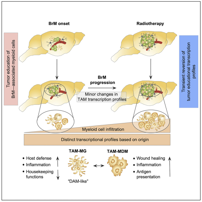
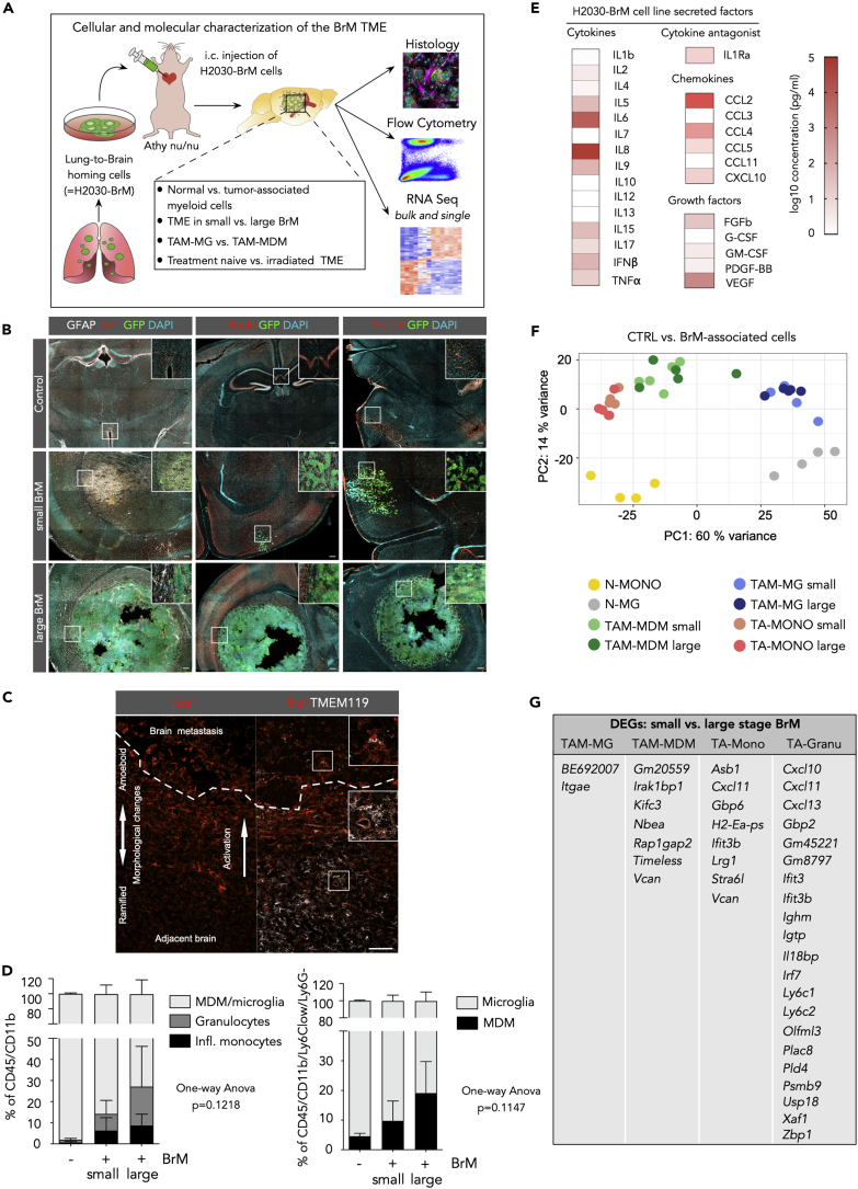
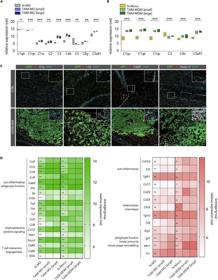
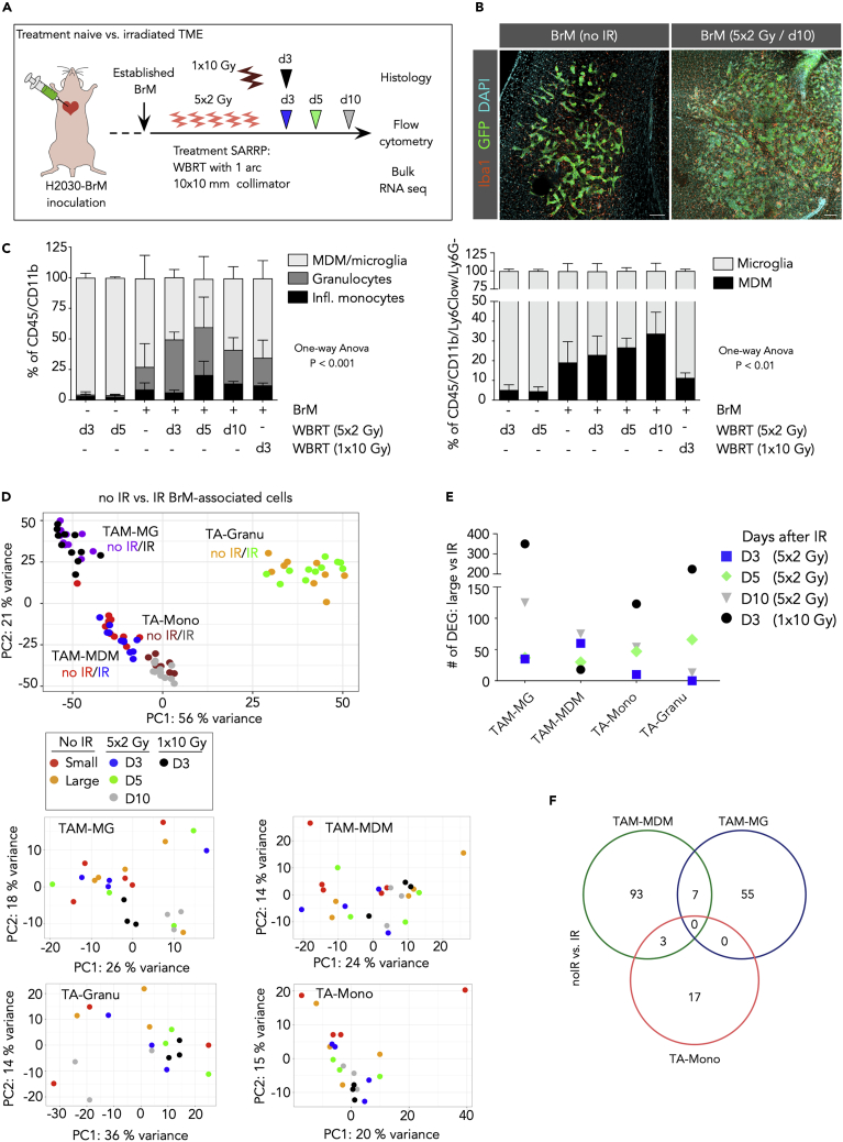
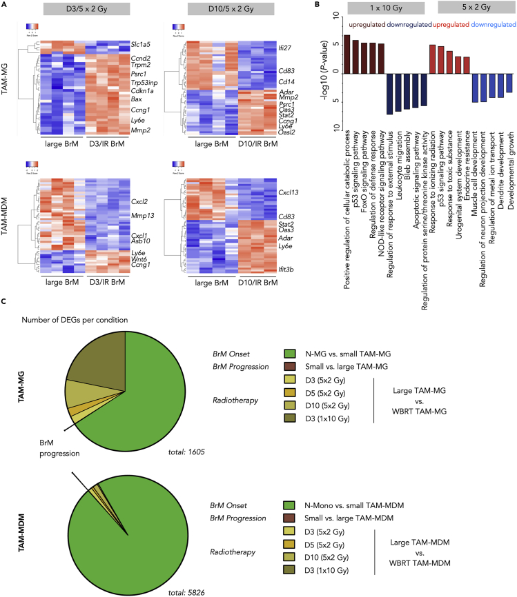
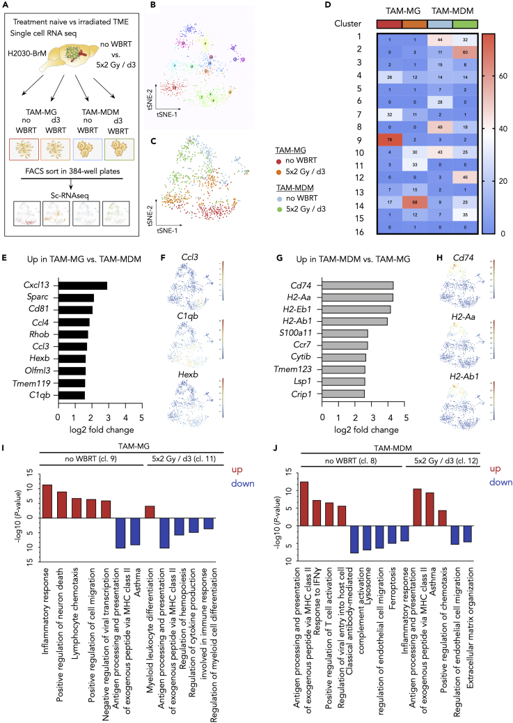
Brain-resident microglia and bone marrow-derived macrophages represent the most abundant non-cancerous cells in the brain tumor microenvironment with critical functions in disease progression and therapeutic response. To date little is known about genetic programs that drive disease-associated phenotypes of microglia and macrophages in brain metastases. Here we used cytometric and transcriptomic analyses to define cellular and molecular changes of the myeloid compartment at distinct stages of brain metastasis and in response to radiotherapy. We demonstrate that genetic programming of tumor education in myeloid cells occurs early during metastatic onset and remains stable throughout tumor progression. Bulk and single cell RNA sequencing revealed distinct gene signatures in brain-resident microglia and blood-borne monocytes/macrophages during brain metastasis and in response to therapeutic intervention. Our data provide a framework for understanding the functional heterogeneity of brain metastasis-associated myeloid cells based on their origin.

Keywords: Immunology; Systems Biology; Transcriptomics.


Conflict of interest statement

Declaration of Interests The authors declare no conflict of interest.

Figures

None
Graphical abstract
Figure 1
Figure 1
Cellular Composition of the Tumor Microenvironment of H2030-BrM (A) Schematic overview of the experimental design and methodology. (B) Representative immunofluorescence images of tumor-free control brain and small and large metastatic lesions in the H2030-BrM model stained with indicated markers. Scale bar, 200 μm. (C) Representative immunofluorescence image of a brain metastatic lesion and adjacent normal brain parenchyma stained for Iba1 and Tmem119. Dotted line depicts the tumor margin. Overview is split into Iba1 staining (left panel) and an overlay of Iba1 and TMEM119 staining (right panel). Scale bar, 100 μm. (D) Quantification of myeloid cell populations in normal brain and in the H2030-BrM model by flow cytometry (n = 3 for each condition). p values were obtained by one-way ANOVA. (E) Quantification of the concentration of secreted factors in tumor cell-conditioned media from H2030BrM cells in vitro (n = 3 replicates). (F) Principle component analysis (PCA) of different cell types in tumor-free versus BrM condition. N-MG, normal microglia; N-Mono, normal blood monocytes; TA, tumor-associated cell type. n = 4 for cells from tumor-free mice and n = 9 for cells isolated from BrM. (G) DEGs in small versus large BrM in the individual cell types. n = 4 for tumor-free mice, n = 4–5 for mice with small BrM, and n = 5 for mice with large BrM.
Figure 2
Figure 2
Tumor Education Signatures of TAMs in BrM (A) Euler diagrams depict the number of unique and shared upregulated (left panel) or downregulated (right panel) DEGs in control versus TAM-MG, or TAM-MDM, or TA-Mono. (B–D) Volcano plot of control cells versus BrM-associated cells in (B) microglia, (C) MDM, and (D) monocytes. Cutoffs: ± 1 log2 fold change and adjusted p value <0.05. (E–G) Functional gene annotation of altered cellular pathways in (E) TAM-MG, (F) TAM-MDM, and (G) TA-Mono compared with control cell types from tumor-free animals. Cutoffs: ± 1, log2 fold change and adjusted p value <0.05. All DEGs or top 3,000 DEGs based on adjusted p values were subjected to analysis. (H–J) Unsupervised clustering of the top 50 DEGs in control versus BrM-associated (small and large stage) TAMs for (H) microglia, (I) MDM, and (J) monocytes. n = 4–5 replicates for each condition.
Figure 3
Figure 3
The Complement System in TAMs and Their Polarization States in BrM (A) Expression of complement members in TAM-MG (B) Expression of complement members in TAM-MDM; for (A) and (B): Values are based on variance-stabilized data (vst (≈log2)) of RNA-seq data. N-Mono: n = 4, N-MG: n = 4, small BrM-associated cells, all other groups: n = 5. Significance based on padj. (C) Representative immunofluorescence images of control and H2030-BrM sections stained for the indicated markers. Scale bars, 100 and 25 μm. (D) Heatmaps display the relative expression level of different activation/polarization marker within both TAM populations. Genes are grouped into M1-like (green, left panel) and M2-like (red, right panel). Values display the average per group based on vst values from the comparison of control (N-MG or N-Mono with n = 4 for each group) versus BrM-associated TAM-MG or TAM-MDM (n = 10 for each group). Significance based on padj.
Figure 4
Figure 4
Effects of Whole-Brain Radiotherapy on the Myeloid Cell Compartment in BrM (A) Schematic representation of the application of whole-brain radiotherapy (WBRT) in two different radiation regimens, 5 × 2 and 1 × 10 Gy. (B) Representative immunofluorescence images of control and irradiated H2030-BrM stained for the indicated markers. Scale bars, 100 μm. (C) Relative quantification of myeloid cell populations in normal brain at d3 and 5 after 5 × 2 Gy WBRT and in H2030-BrM at d3, d5, and d10 after 5 × 2 Gy WBRT or d3 after 1 × 10 Gy compared with untreated BrM (n = 3 for each condition). Data are represented as mean ± SD. p values were obtained by one-way ANOVA. (D) PCA plot shows clustering of individual samples (all non-irradiated versus all irradiated samples) indicating that clustering is mostly driven by cell type with minor effect of IR. PCA plots per cell type indicate heterogeneous clustering across conditions (see small box). (E) Amount of significant DEGs (cutoff: BM > 20, adj. p value <0.05) in different cell types, by comparing RNA-seq data of large stage BrM-associated samples (n = 5) with its irradiated BrM-associated cellular counterparts at different days upon WBRT (5 × 2 Gy/d3 and d5 n = 4, 1 × 10 Gy/d3 and 5 × 2 Gy/d10 n = 3). (F) Venn diagram depicts shared and unique DEGs (cutoff: BM > 20, adj. p value <0.05) in each cell type from RNA-seq data of H2030-BrM-associated immune cells, comparing all non-irradiated (no IR) versus all irradiated (IR) groups; note: granulocytes did not reveal any DEG (5 × 2 Gy/d3 and d5 n = 4, 1 × 10 Gy/d3 and 5 × 2 Gy/d10 n = 3).
Figure 5
Figure 5
Effects of Whole-Brain Radiotherapy on Distinct TAM populations in BrM (A) Unsupervised hierarchical clustering heatmaps depicting Top 50 DEGs in TAM-MG (upper panel) and TAM-MDM (lower panel) of large stage BrM (each n = 4) versus d3 (left, both cell types n = 4) or d10 (right, both cell types n = 3), respectively. (B) Top five functional gene annotation of DEG in the indicated conditions (cutoff: BM > 20, adj. p value <0.05; log2 fold change). (C) Pie chart depicting the amount of significantly DEGs in different comparisons of distinct MG or MDM conditions, reflecting BrM onset, progression, and radiotherapy, respectively. Cutoffs: BM > 20, adj. p value < 0.05. n = 3–5 per condition.
Figure 6
Figure 6
Single-Cell RNA-Seq Reveals Heterogeneous TAM Populations (A) Schematic overview of the experimental design for the single-cell RNA-seq analyses. (B) tSNE plot of all analyzed cells reveal the existence of 16 clusters within the four analyzed experimental groups (untreated TAM-MG, 5 × 2 Gy/d3 TAM-MG, untreated TAM-MDM, 5 × 2 Gy/d3 TAM-MDM). (C) tSNE plot of all analyzed cells with color coding for each experimental condition indicates representation of individual conditions in multiple cluster. (D) Heatmap depicts the number of cells per condition contributing to the individual cluster. (E) Top 10 upregulated genes in TAM-MG versus TAM-MDM based on log2 fold change. (F) tSNE plots indicate the expression of representative genes in TAM-MG cluster. (G) Top 10 upregulated genes in TAM-MDM versus TAM-MG based on log2 fold change. (H) tSNE plots indicate the expression of representative genes in TAM-MDM cluster. (I and J) Functional gene annotation of up- (red) and downregulated (blue) genes for (I) untreated TAM-MG (cl.9) versus 5 × 2Gy/d3 TAM-MG (cl.11) and (J) untreated TAM-MDM (cl.8) versus (cl.12) 5 × 2Gy/d3 TAM-MDM. A maximum of five pathways is represented.

References

    1. Ajami B., Samusik N., Wieghofer P., Ho P.P., Crotti A., Bjornson Z., Prinz M., Fantl W.J., Nolan G.P., Steinman L. Single-cell mass cytometry reveals distinct populations of brain myeloid cells in mouse neuroinflammation and neurodegeneration models. Nat. Neurosci. 2018;21:541–551. - PMC - PubMed
    1. Albulescu R., Codrici E., Popescu I.D., Mihai S., Necula L.G., Petrescu D., Teodoru M., Tanase C.P. Cytokine patterns in brain tumour progression. Mediators Inflamm. 2013;2013:979748. - PMC - PubMed
    1. Bennett M.L., Bennett F.C., Liddelow S.A., Ajami B., Zamanian J.L., Fernhoff N.B., Mulinyawe S.B., Bohlen C.J., Adil A., Tucker A. New tools for studying microglia in the mouse and human CNS. Proc. Natl. Acad. Sci. U S A. 2016;113:E1738–E1746. - PMC - PubMed
    1. Betancur P.A., Abraham B.J., Yiu Y.Y., Willingham S.B., Khameneh F., Zarnegar M., Kuo A.H., McKenna K., Kojima Y., Leeper N.J. A CD47-associated super-enhancer links pro-inflammatory signalling to CD47 upregulation in breast cancer. Nat. Commun. 2017;8:14802. - PMC - PubMed
    1. Bouwens van der Vlis T.A.M., Kros J.M., Mustafa D.A.M., van Wijck R.T.A., Ackermans L., van Hagen P.M., van der Spek P.J. The complement system in glioblastoma multiforme. Acta Neuropathol. Commun. 2018;6:91. - PMC - PubMed