Review
. 2020 Aug:63:154-161.
doi: 10.1016/j.conb.2020.04.005. Epub 2020 May 29.

Mining the jewels of the cortex's crowning mystery

Affiliations
Review

Mining the jewels of the cortex's crowning mystery

Leena A Ibrahim et al. Curr Opin Neurobiol. 2020 Aug.

Abstract

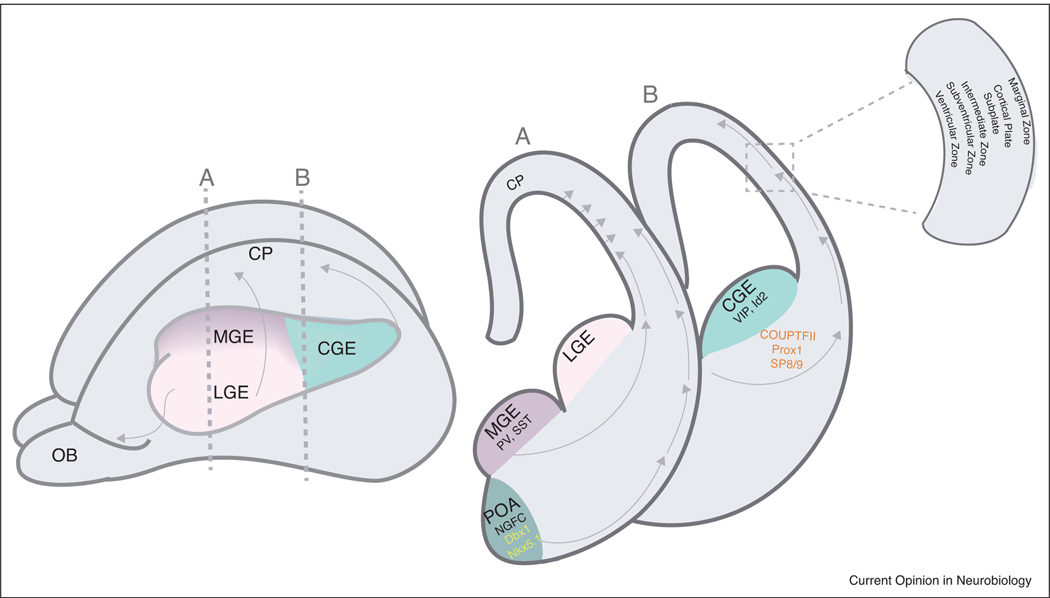
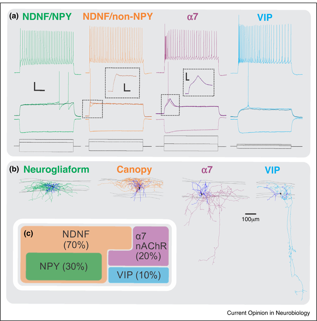
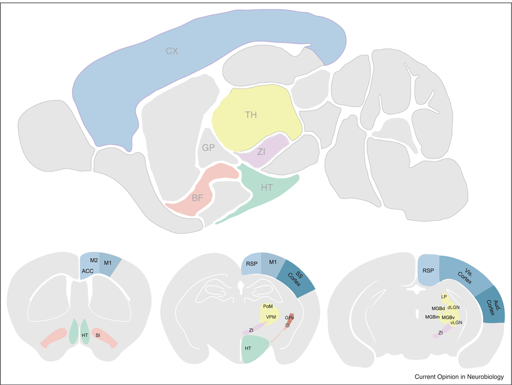
Neocortical Layer 1 consists of a dense mesh of excitatory and inhibitory axons, dendrites of pyramidal neurons, as well as neuromodulatory inputs from diverse brain regions. Layer 1 also consists of a sparse population of inhibitory interneurons, which are appropriately positioned to receive and integrate the information from these regions of the brain and modulate cortical processing. Despite being among the sparsest neuronal population in the cortex, Layer 1 interneurons perform powerful computations and have elaborate morphologies. Here we review recent studies characterizing their origin, morphology, physiology, and molecular profiles, as well as their connectivity and in vivo response properties.


Conflict of interest statement

Conflict of interest statement

Nothing declared.

Figures

Figure 1
Figure 1
Developmental origin of Layer 1 interneurons. Left, schematic diagram of an embryonic mouse brain (~E14) highlighting the ganglionic eminences from which inhibitory interneurons are derived. Medial ganglionic eminence (MGE) gives rise to PV and SST interneurons, Lateral ganglionic eminence (LGE) gives rise to the interneurons of the olfactory bulb (OB); and Caudal ganglionic eminence (CGE) gives rise to the VIP and Id2 interneuron populations. Vertical dashed lines indicate the levels of the coronal sections in the right panel. Right panel, coronal sections through the three eminences as well as the preoptic area (POA). Layer 1 interneurons are derived from the CGE and POA. Arrows indicate the routes of migration that the interneurons prefer to reach the cortical plate. Boxed inset highlights the structures surrounding the cortical plate present at this embryonic age.
Figure 2
Figure 2
Subtypes of Layer 1 interneurons. (a) Electrophysiological properties of the four different L1 subtypes: NDNF/NPY positive; NDNF/NPY negative, VIP, and alpha-7. Middle panel illustrates the response of the neurons to a threshold current injection. Notice that the NDNF/NPY positive population has a late spiking property, and the alpha-7 population has a depolarizing hump near threshold. (b) The morphologies associated with the four subtypes. Notice the elaborate axonal and dendritic arborization of the neurogliaform and the canopy cells (NDNF population) mostly restricted to Layer 1; whereas the alpha-7 and VIP possess a descending axon projecting down to deeper layers. (c) Relative proportions of the four interneuron subtypes in Layer 1.
Figure 3
Figure 3
Connectivity of Layer 1 interneurons. Upper panel, saggital section of an adult mouse brain highlighting the relevant brain regions that project to Layer 1. Thalamus (TH), Cortex (CX), Zona Incerta (ZI), Basal Forebrain (BF), Hypothalamus (HT). Bottom panels, coronal sections detailing the structures in the saggital section above. Cg (cingulate cortex), M2 (premotor cortex), M1 (motor cortex), SI (substantia innominata; part of BF), RSP (retrosplenial cortex), Som Cx (Somatosensory cortex), LP (Lateral posterior nucleus), dLGN (dorsal lateral geniculate nucleus), vLGN (ventral lateral geniculate nucleus), MGB (medial geniculate nucleus). MGB consists of d (dorsal) m (medial) and v (ventral) subdivisions. Note: These highlighted structures illustrate a general pattern of connectivity of Layer 1 interneurons in the sensory cortices. For example, Layer 1 interneurons in Som cortex receive projections from M1, M2, Cg, RSP, and BF, as well as their respective thalamic nuclei PO and VPM. In the visual cortex, L1 interneurons also receive inputs from M1, M2, Cg, RSP and BF, as well as their respective thalamic nuclei, LGN, and LP. Also note, this is not an exhaustive list of structures projecting to L1 interneurons in specific brain regions but a general pattern observed across multiple areas of the sensory cortex.

References

    1. Rudy B, Fishell G, Lee S, Hjerling-Leffler J: Three groups of interneurons account for nearly 100% of neocortical GABAergic neurons. Dev Neurobiol 2011, 71:45–61. - PMC - PubMed
    1. Hubel DH: Cortical neurobiology: a slanted historical perspective. Annu Rev Neurosci 1982, 5:363–370. - PubMed
    1. Xu Q, Cobos I, De La Cruz E, Rubenstein JL, Anderson SA: Origins of cortical interneuron subtypes. J Neurosci 2004, 24:2612–2622. - PMC - PubMed
    1. Butt SJ, Fuccillo M, Nery S, Noctor S, Kriegstein A, Corbin JG, Fishell G: The temporal and spatial origins of cortical interneurons predict their physiological subtype. Neuron 2005, 48:591–604. - PubMed
    1. Yozu M, Tabata H, Nakajima K: The caudal migratory stream: a novel migratory stream of interneurons derived from the caudal ganglionic eminence in the developing mouse forebrain. J Neurosci 2005, 25:7268–7277. - PMC - PubMed

Publication types