Colorectal cancer with invasive micropapillary components (IMPCs) shows high lymph node metastasis and a poor prognosis: A retrospective clinical study
- PMID: 32481300
- PMCID: PMC7249862
- DOI: 10.1097/MD.0000000000020238
Colorectal cancer with invasive micropapillary components (IMPCs) shows high lymph node metastasis and a poor prognosis: A retrospective clinical study
Abstract
Objects: The present study aimed to identify the clinicopathological characteristics of colorectal cancer (CRC) with invasive micropapillary components (IMPCs) and the relationship between different amounts of micropapillary components and lymph node metastasis.
Methods: A cohort of 363 patients with CRC who underwent surgical treatment in the Second Affiliated Hospital of Zhejiang University School of Medicine between January 2013 and December 2016 were retrospectively reviewed. We compared the clinicopathological characteristics, including survival outcomes and immunohistochemical profiles (EMA, MUC1, MLH1, MSH2, MSH6, and PMS2), between CRC with IMPCs and those with conventional adenocarcinoma (named non-IMPCs in this study). Logistic regression was used to identify the association between IMPCs and lymph node invasion. A multivariate analysis was performed using the Cox proportional hazard model to evaluate significant survival predictors.
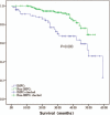
Results: Among 363 patients, 76 cases had IMPCs, including 22 cases with a lower proportion of IMPCs (≤5%, IMPCs-L) and 54 cases with a higher proportion (>5%, IMPCs-H). Compared to the non-IMPC group, the IMPC group (including both IMPC-L and IMPC-H) had a lower degree of tumor differentiation (P = .000), a higher N-classification (P = .000), more venous invasion (P = .019), more perineural invasion (P = .025) and a later tumor node metastasis (TNM) stage (P = .000). Only tumor differentiation (P = .031) and tumor size (P = .022) were different between IMPCs-L and IMPCs-H. EMA/MUC1 enhanced the characteristic inside-out staining pattern of IMPCs, whereas non-IMPCs showed luminal staining patterns. The percentage of mismatch repair deficiency (dMMR) in the non-IMPC group was much higher than that in the IMPC group (14.7% vs 4.7%). The overall survival time of patients with IMPCs was significantly less than that of patients with non-IMPCs (P = .002), then that of IMPCs-H was lower than that of IMPCs-L (P = .030). Logistic regression revealed that patients with IMPCs were associated with lymph metastasis, regardless of the proportion of IMPCs. Multivariate analysis demonstrated both IMPCs-L and IMPCs-H as negative prognostic factors.
Conclusions: IMPCs are significantly associated with lymph node metastasis and poor outcome, and even a minor component (≤5%) may render significant information and should therefore be part of the pathology report.
Conflict of interest statement
The authors declare no conflict of interest.
Figures


References
-
- Siriaunkgul S, Tavassoli FA. Invasive micropapillary carcinoma of the breast. Mod Pathol 1993;6:660–2. - PubMed
-
- Amin MB, Ro JY, el-Sharkawy T, et al. Micropapillary variant of transitional cell carcinoma of the urinary bladder. Histologic pattern resembling ovarian papillary serous carcinoma. Am J Surg Pathol 1994;18:1224–32. - PubMed
-
- Amin MB, Tamboli P, Merchant SH, et al. Micropapillary component in lung adenocarcinoma: a distinctive histologic feature with possible prognostic significance. Am J Surg Pathol 2002;26:358–64. - PubMed
-
- Smith Sehdev AE, Sehdev PS, Kurman RJ. Noninvasive and invasive micropapillary (low-grade) serous carcinoma of the ovary: a clinicopathologic analysis of 135 cases. Am J Surg Pathol 2003;27:725–36. - PubMed
-
- Michal M, Skálová A, Mukensnabl P. Micropapillary carcinoma of the parotid gland arising in mucinous cystadenoma. Virchows Arch 2000;437:465–8. - PubMed
MeSH terms
Substances
Supplementary concepts
LinkOut - more resources
Full Text Sources
Medical
Research Materials
Miscellaneous

