Review
. 2020 May 28;11(6):595.
doi: 10.3390/genes11060595.

Epigenetic Factors That Control Pericentric Heterochromatin Organization in Mammals

Affiliations
Review

Epigenetic Factors That Control Pericentric Heterochromatin Organization in Mammals

Salvatore Fioriniello et al. Genes (Basel). .

Abstract

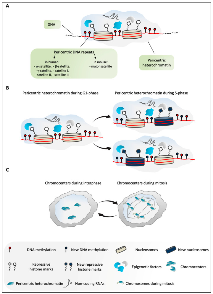
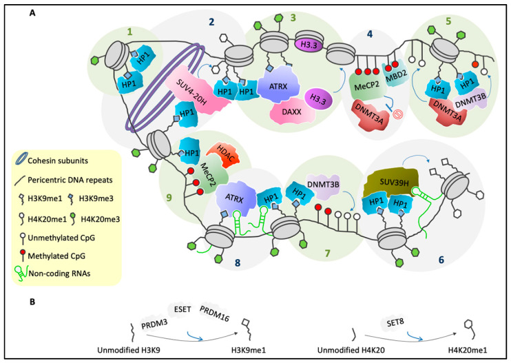
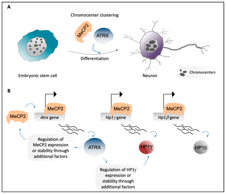
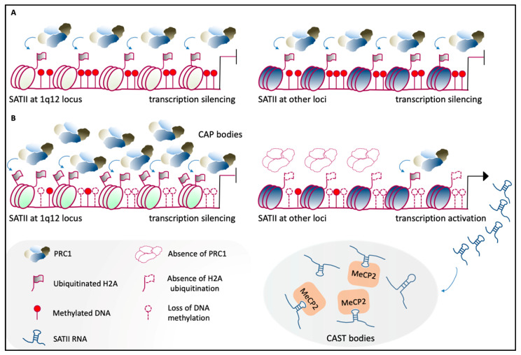
Pericentric heterochromatin (PCH) is a particular form of constitutive heterochromatin that is localized to both sides of centromeres and that forms silent compartments enriched in repressive marks. These genomic regions contain species-specific repetitive satellite DNA that differs in terms of nucleotide sequences and repeat lengths. In spite of this sequence diversity, PCH is involved in many biological phenomena that are conserved among species, including centromere function, the preservation of genome integrity, the suppression of spurious recombination during meiosis, and the organization of genomic silent compartments in the nucleus. PCH organization and maintenance of its repressive state is tightly regulated by a plethora of factors, including enzymes (e.g., DNA methyltransferases, histone deacetylases, and histone methyltransferases), DNA and histone methylation binding factors (e.g., MECP2 and HP1), chromatin remodeling proteins (e.g., ATRX and DAXX), and non-coding RNAs. This evidence helps us to understand how PCH organization is crucial for genome integrity. It then follows that alterations to the molecular signature of PCH might contribute to the onset of many genetic pathologies and to cancer progression. Here, we describe the most recent updates on the molecular mechanisms known to underlie PCH organization and function.

Keywords: ATRX; DNA methylation; HP1; MeCP2; non-coding RNAs; pericentric heterochromatin; repressive compartments; satellite DNA.


Conflict of interest statement

The authors declare that they have no conflicts of interest.

Figures

Figure 1
Figure 1
(A) Overview of pericentric heterochromatin (PCH) in mammals. PCH is constituted by highly methylated pericentric DNA repeats (α-, β-, and γ-satellites, satellites I, II, III in humans; major satellite in mice) [9,16]. It is enriched in several epigenetic factors, non-coding RNAs, and repressive histone modifications [4,13]. (B) Schematic representation of PCH in G1 and S phases of the cell cycle in mammals. According to the current model, during DNA replication, PCH is assembled through the incorporation of both old and newly synthesized histones. Similarly, epigenetic marks are enriched at PCH, including DNA methylation and repressive histone modifications, and these are inherited from the parental structure and/or established ex novo by different epigenetic factors. These processes ensure faithful maintenance of the PCH structure and its repressive environment during the cell cycle [4,13]. (C) In murine cells in interphase, PCH of different chromosomes is organized in highly compacted structures, termed chromocenters. During mitosis, the dissociation of the chromocenters into individual chromosomes takes place [4,16].
Figure 2
Figure 2
Several factors are involved in PCH organization. (A) Schematic representation of the general molecular structure of mammalian PCH during interphase. Step 1: HP1s bind to H3K9me3 and can self-interact [44]. Step 2: HP1s recruit the SUV4-20H enzymes, which can convert H4K20me1 into H4K20me3. Moreover, SUV4-20H binds cohesin subunits as well as HP1s, which reinforces chromatin compaction [25,45]. Step 3: HP1s and H3K9me3 provide a binding platform for ATRX [46,47], which in complex with DAXX mediates deposition of H3.3 [48]. Step 4: MeCP2 and MBD2 can form heterodimers [49], and they bind methylated CpGs [50]. Moreover, MeCP2 recruits DNMT3A and maintains it in a reversible inactive state [51]. Step 5: HP1s recruit DNMT3A and DNMT3B [16,40], which catalyze methylation of CpGs [52] and can form heterodimers [40]. Step 6: HP1–HP1 dimers recruit the SUV39H enzymes [39] that can trimethylate H3K9me1 on adjacent nucleosomes [16,31]. SUV39H binding to PCH is stabilized by an RNA component [53]. Trimethylation of H4K20me1 requires pre-existing H3K9me3 and HP1s [25]. Step 7: HP1s recruit DNMT3B, which then methylates CpGs [40,52]. Step 8: Accumulation of HP1 [54] and ATRX [20] at PCH also requires an RNA component. Step 9: ATRX binds MeCP2 [55,56], which then recruits a complex that has histone deacetylase activity [14]. Furthermore, MeCP2 interacts with HP1s [57]. (B) PRDM3, ESET, and PRDM16 promote the conversion of unmethylated H3K9 into H3K9me1 [32] (left). SET8 catalyzes the monomethylation of H4K20 tails [58,59] (right).
Figure 3
Figure 3
(A) Embryonic stem cells show high numbers of chromocenters per nucleus (left). In neurons, chromocenters increase in size and decrease in number due to aggregation of PCH of different chromosomes (chromocenter clustering) [18,20] (right). MeCP2 [18] and ATRX [20] are important players in chromocenter clustering during neural differentiation. (B) MeCP2 directly promotes expression of genes that encode PCH-associated factors, including Atrx, Hp1γ, and Hp1β (top). ATRX regulates expression and/or stability of MeCP2 and HP1γ, probably through the involvement of additional factors [20] (bottom).
Figure 4
Figure 4
Human PCH organization in health and disease. (A) Under normal physiological conditions, satellite II (SATII) at the 1q12 locus (left) and at other chromosomal loci (right) is highly methylated at the DNA level and binds the PRC1 complex, which mediates H2A ubiquitination. This molecular landscape leads to the maintenance of the transcriptionally inactive state [151]. (B) In cancer cells, the loss of methylation across SATII loci causes hyper-accumulation of PRC1 proteins at SATII at the 1q12 locus, which leads to the formation of cancer-associated polycomb (CAP) bodies (top-left). This mechanism maintains the silencing of 1q12-SATII, which is reinforced by increased H2A ubiquitination. At other loci, SATII shows less accumulation of ubiquitinated H2A, and becomes transcriptionally active (top right). This leads to the formation of cancer-associated satellite transcript (CAST) bodies, in which there is an aberrant accumulation of SATII RNAs. These aggregates sequester epigenetic factors, including MeCP2. Adapted from [151].

References

    1. O’Keefe R.T., Henderson S.C., Spector D.L. Dynamic organization of DNA replication in mammalian cell nuclei: Spatially and temporally defined replication of chromosome-specific alpha-satellite DNA sequences. J. Cell Biol. 1992;116:1095–1110. doi: 10.1083/jcb.116.5.1095. - DOI - PMC - PubMed
    1. Kim S.M., Dubey D.D., Huberman J.A. Early-replicating heterochromatin. Genes Dev. 2003;17:330–335. doi: 10.1101/gad.1046203. - DOI - PMC - PubMed
    1. Hollo G., Kereso J., Praznovszky T., Cserpan I., Fodor K., Katona R., Csonka E., Fatyol K., Szeles A., Szalay A.A., et al. Evidence for a megareplicon covering megabases of centromeric chromosome segments. Chromosome Res. 1996;4:240–247. doi: 10.1007/BF02254965. - DOI - PubMed
    1. Almouzni G., Probst A.V. Heterochromatin maintenance and establishment: Lessons from the mouse pericentromere. Nucleus. 2011;2:332–338. doi: 10.4161/nucl.2.5.17707. - DOI - PubMed
    1. Brown S.W. Heterochromatin. Science. 1966;151:417–425. doi: 10.1126/science.151.3709.417. - DOI - PubMed

Publication types

LinkOut - more resources