

Choline PET/CT in Multiple Myeloma
- PMID: 32481661
- PMCID: PMC7352763
- DOI: 10.3390/cancers12061394
Choline PET/CT in Multiple Myeloma
Abstract
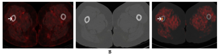
The field of multiple myeloma (MM) imaging has evolved. The International Myeloma Working Group recently recommended performing 18F-fluorodeoxyglucose glucose (18FDG) positron emission tomography/computed tomography (PET/CT) with the aim of staging MM patients at baseline and evaluating response to therapy. Novel oncological radiotracers such as 11C-Choline and 18F-Fluorocholine, have been studied in comparison with 18FDG, mostly in MM patients presenting with refractory disease or suspected relapse. Choline-based tracers may overcome some limitations of 18FDG, which include a lack of sensitivity in depicting skull lesions and the fact that 10% of MM patients are FDG-negative. The majority of MM lesions display a higher uptake of Choline than FDG. Also, in many situations, Choline may offer better lesion visualization, with a higher tumor to background ratio; however, various patterns of Choline and FDG uptake have been observed in MM and some limitations, notably as regards liver lesions, should be recognized. Overall, Choline may provide additional detection of up to 75% more lesions. This article aims to provide a comprehensive review of the potential role of Choline in multiple myeloma, as compared to FDG, encompassing Choline physiopathology as well as data from clinical studies.
Keywords: 11C-Choline; 18F-Choline; 18FDG; PET/CT; multiple myeloma.
Conflict of interest statement
The authors declare no conflicts of interest.
Figures
References
-
- Rajkumar S.V., Dimopoulos M.A., Palumbo A., Blade J., Merlini G., Mateos M.V., Kumar S., Hillengass J., Kastritis E., Richardson P., et al. International Myeloma Working Group updated criteria for the diagnosis of multiple myeloma. Lancet Oncol. 2014;15:e538–e548. doi: 10.1016/S1470-2045(14)70442-5. - DOI - PubMed
-
- Cavo M., Terpos E., Nanni C., Moreau P., Lentzsch S., Zweegman S., Hillengass J., Engelhardt M., Usmani S.Z., Vesole D.H., et al. Role of (18)F-FDG PET/CT in the diagnosis and management of multiple myeloma and other plasma cell disorders: A consensus statement by the International Myeloma Working Group. Lancet Oncol. 2017;18:e206–e217. doi: 10.1016/S1470-2045(17)30189-4. - DOI - PubMed
-
- Hillengass J., Usmani S., Rajkumar S.V., Durie B.G.M., Mateos M.V., Lonial S., Joao C., Anderson K.C., Garcia-Sanz R., Riva E., et al. International myeloma working group consensus recommendations on imaging in monoclonal plasma cell disorders. Lancet Oncol. 2019;20:e302–e312. doi: 10.1016/S1470-2045(19)30309-2. - DOI - PubMed
-
- DeGrado T.R., Coleman R.E., Wang S., Baldwin S.W., Orr M.D., Robertson C.N., Polascik T.J., Price D.T. Synthesis and evaluation of 18F-labeled choline as an oncologic tracer for positron emission tomography: Initial findings in prostate cancer. Cancer Res. 2001;61:110–117. - PubMed
Publication types
LinkOut - more resources
Full Text Sources
