Review
. 2020 Jun;20(6):e117-e128.
doi: 10.1016/S1473-3099(20)30148-1. Epub 2020 May 5.

Cavitary tuberculosis: the gateway of disease transmission

Affiliations
Review

Cavitary tuberculosis: the gateway of disease transmission

Michael E Urbanowski et al. Lancet Infect Dis. 2020 Jun.

Abstract

Tuberculosis continues to be a major threat to global health. Cavitation is a dangerous consequence of pulmonary tuberculosis associated with poor outcomes, treatment relapse, higher transmission rates, and development of drug resistance. However, in the antibiotic era, cavities are often identified as the most extreme outcome of treatment failure and are one of the least-studied aspects of tuberculosis. We review the epidemiology, clinical features, and concurrent standards of care for individuals with cavitary tuberculosis. We also discuss developments in the understanding of tuberculosis cavities as dynamic physical and biochemical structures that interface the host response with a unique mycobacterial niche to drive tuberculosis-associated morbidity and transmission. Advances in preclinical models and non-invasive imaging can provide valuable insights into the drivers of cavitation. These insights will guide the development of specific pharmacological interventions to prevent cavitation and improve lung function for individuals with tuberculosis.


Conflict of interest statement

Declaration of interests

The authors declare no conflicts of interest.

Figures
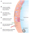
Figure 1.
Figure 1.
Why is cavitary TB so hard to treat? (A) High concentrations of extracellular bacteria grow in the loose necrotic debris at the interior surface of the cavity. (B) The bacterial proliferation leads to replication induced mutations at drug-resistance determining loci and a high probability of mutants with acquired drug resistance., (C) Extracellular collagen matrix is depleted within caseous lesions and in the cavity wall. Depletion of extracellular collagen matrix facilitates the formation and growth of cavities since the remaining necrotic debris are easily evacuated through an adjoining bronchus. Once depleted, the healing response is unable to regenerate the basement membrane or lung tissue. Individuals face lifelong pulmonary deficits and a high-risk for opportunistic infections within persistent lung cavities. (D) The inner layer of the cavity wall is composed of necrotic debris. Few immune cells penetrate this region to aid in control over M. tuberculosis replication, and this contributes to the high bacterial burden. (E) Vascular necrosis around the cavity and strong drug-binding properties of caseum result in poor anti-mycobacterial drug penetration which also contributes to the high bacterial burden.,, The effects of sub-optimal drug penetration may also drive selection for drug-resistant mutants. (F) Cavities often persist even after they are sterilized of mycobacteria and are replaced with scar tissue (closed healing). Therefore, cavitation can lead to loss of lung volume and chronic pulmonary deficits. If the cavities persist following curative therapy (open healing), then they become a vulnerable environment for secondary colonization by opportunistic infections. The combination of warm temperatures, high humidity, immune-sheltering, and lack of innate-defenses provide an opportunity for secondary colonization, often by Aspergillus spp.
Figure 2.
Figure 2.
The architecture of TB cavities. (A) A transverse lung field CT-scan reconstruction from a TB patient showing a large cavity. (B) The bronchopulmonary segment distribution of 287 cavities from the TB Portals Program database are represented as a heatmap of the % of total cavities evaluated. (C) Location and size of cavities analyzed in B. (D) A gross image of a large apical cavity from a TB patient. The image was obtained from the autopsy record of the Johns Hopkins Hospital and used with permission of the Johns Hopkins University Chesney Medical Archives. (E) Histology of the cavity wall of an immunocompetent human patient with pulmonary TB. Each high magnification image is a serial section from the field identified by the box in the low magnification image of the entire cavity. Data for A-C were obtained from the TB Portals, which is an open-access TB data resource supported by the National Institute of Allergy and Infectious Diseases (NIAID) Office of Cyber Infrastructure and Computational Biology (OCICB). These data were collected and submitted by members of the TB Portals Consortium. Investigators and other data contributors that originally contributed the data to the TB Portals did not participate in the design or analysis of this study.
Figure 3.
Figure 3.
Radiologic and histologic examples of cavities from cavitary TB models. Cavities in animal models infected with M. tuberculosis have similar radiological and histological characteristics to those found in humans. (A) Common marmoset (adapted with permission from Via et al.) (B) New Zealand white rabbit. (C) C3HeB/FeJ mouse (adapted with permission from Ordonez et al.).
Figure 4.
Figure 4.
Overview of the drivers of pulmonary cavitation in TB. (A) The biochemical drivers of cavitation cause basement membrane destruction and pathologic fibrosis. Black arrows indicate extracellular signaling pathways. ECM = extracellular matrix. (B) The biophysical drivers of cavitation. The prefix ‘p’ indicates the partial pressure of gases in each type of cavity compartment. Black arrows denote the bulk flow of air due to respiratory motion and/or influence by caseous occlusion (yellow material). (C) A possible model outlining the microbial drivers of cavitation. The model is based heavily on Yamamura and colleagues’ seminal work on the cavity-inducing constituents of the heat-inactivated bacillus. Figure S5 provides an overview of Yamamura’s experiments. (+) indicates that the chemical constituent has a role in increasing a given process. (−) indicates that the chemical constituent has a role in decreasing a given process. (D) The immunological profile of cavitary TB in the bronchoalveolar lavage/sputum compartment and the peripheral blood compartment. All comparisons are from studies comparing individuals with cavitary TB to individuals with non-cavitary TB. ↑ indicates an increased concentration of cells or cytokines. ↓ indicates a decreased concentration of cells or cytokines. ↔ indicates no difference observed between groups. Double arrows (e.g. ↑↑) indicate a strong trend. Table S2 shows the magnitude of these trends with study citations.

References

    1. Global tuberculosis report 2019. Geneva: World Health Organization, 2019.
    1. Benator D, Bhattacharya M, Bozeman L, et al. Rifapentine and isoniazid once a week versus rifampicin and isoniazid twice a week for treatment of drug-susceptible pulmonary tuberculosis in HIV-negative patients: a randomised clinical trial. Lancet 2002; 360(9332): 528–34. - PubMed
    1. Palaci M, Dietze R, Hadad DJ, et al. Cavitary disease and quantitative sputum bacillary load in cases of pulmonary tuberculosis. Journal of clinical microbiology 2007; 45(12): 4064–6. - PMC - PubMed
    1. Dannenberg AM Jr. Pathogenesis of human pulmonary tuberculosis: insights from the rabbit model. Washington: ASM Press; 2006.
    1. Canetti G The tubercle bacillus in the pulmonary lesion of man: histobacteriology and its bearing on the therapy of pulmonary tuberculosis: Springer Publishing Company; 1955.

Publication types

MeSH terms

Substances