Review
. 2020 Jun 1;34(11-12):733-744.
doi: 10.1101/gad.337196.120.

Progress toward understanding chromosome silencing by Xist RNA

Affiliations
Review

Progress toward understanding chromosome silencing by Xist RNA

Neil Brockdorff et al. Genes Dev. .

Abstract

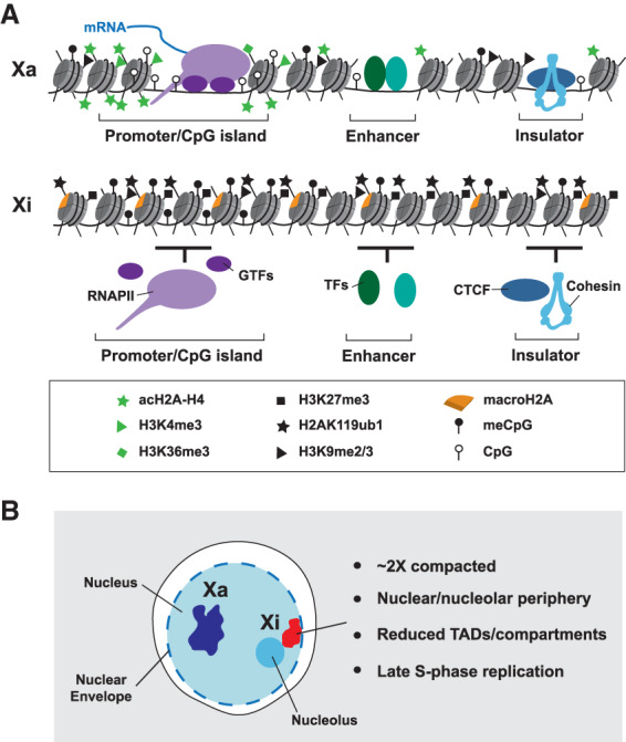
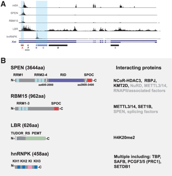
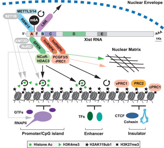
The X inactive-specific transcript (Xist) gene is the master regulator of X chromosome inactivation in mammals. Xist produces a long noncoding (lnc)RNA that accumulates over the entire length of the chromosome from which it is transcribed, recruiting factors to modify underlying chromatin and silence X-linked genes in cis Recent years have seen significant progress in identifying important functional elements in Xist RNA, their associated RNA-binding proteins (RBPs), and the downstream pathways for chromatin modification and gene silencing. In this review, we summarize progress in understanding both how these pathways function in Xist-mediated silencing and the complex interplay between them.

Keywords: LBR; NCoR–HDAC3; Polycomb; RBM15; SPEN; X chromosome inactivation; Xist; chromatin; m6A RNA methylation.


Figures

Figure 1.
Figure 1.
Features of the inactive X chromosome. (A) Comparison of chromatin features on the active X chromosome (Xa) and the inactive X chromosome (Xi) at the nucleosomal scale. Xi is characterized by depletion of histone modifications associated with gene activity, shown here histone acetylation, normally enriched at promoters and enhancers, H3K4me3, enriched at promoters and H3K36me3, high levels of which occur within the bodies of active genes. Additionally, Xi chromatin has enhanced levels of histone modifications associated with gene repression; for example, H3K27me3, H2AK119ub1, H3K9me2/3, and high levels of the histone variant macroH2A. DNA methylation is acquired at CpG islands of Xi genes. RNA polymerase II (RNAPII) and associated general transcription factors (GTFs) are depleted from Xi, as are enhancer-bound transcription factors (TFs), as evidenced by reduced chromatin accessibility seen using ATAC-seq. Similarly, there are reduced levels of the insulator protein CTCF and the cohesin complex at insulator/boundary elements. (B) Changes in higher order structure of the Xi chromosome. Key examples are an approximately twofold chromatin compaction relative to Xa, association of Xi with the nuclear and/or nucleolar periphery, reduced TAD and compartment structure (related to reduced CTCF/cohesin binding), with interactions instead occurring within one of two large megadomains, and synchronous replication across most of the chromosome, usually in late S phase.
Figure 2.
Figure 2.
Xist RNA-binding proteins (RBPs) with a role in chromosome silencing. (A) Binding profiles of key RBPs discussed in this article. iCLIP/eCLIP data obtained from various sources (Chen et al. 2016; Lu et al. 2016; Cirillo et al. 2017; Nesterova et al. 2019) is shown against a map of the Xist gene with tandem repeat elements A–F indicated below. A-repeat and B/C-repeat regions are highlighted with pale-blue shading. The m6A track illustrates sites of m6A deposition as determined by m6A-seq (Coker et al. 2020). Gray bar indicates the location of LBS on Xist. (B) Features of the RBPs SPEN, RBM15, LBR, and hnRNPK. Schematics indicate domain architecture with key interacting partners shown at the right. Validated direct interactors are shown in black. (RRM) RNA recognition motif; (RID) RBPJ interaction domain; (SPOC) Spen paralog and ortholog C-terminal; (RS) arginine/serine; (PEMT) phosphatidylethanolamine N-methyltransferase; (KH) K homology; (KI) K interaction.
Figure 3.
Figure 3.
Models and assays for X inactivation studies. (A) Early mouse embryos. X inactivation (indicated as condensed bar with Xist RNA represented as green dashed line) proceeds at the two- to four-cell stage (E1.5–E2.5), initially on the paternally inherited X chromosome (Xp), referred to as imprinted X chromosome inactivation (iXCI). iXCI is maintained in trophectoderm (orange) and extraembryonic endoderm (green) derived tissues through development (shown here E3.5–E10.5), whereas in cells of the embryo proper (blue), initial reactivation of Xp at E3.5 is followed by random X inactivation (rXCI) of Xp or the maternal X chromosome (Xm) at around E5.5. (B) In XX mouse embryonic stem cells (mESCs) both X chromosomes are active. Random X inactivation proceeds following the onset of cellular differentiation in vitro. (C,D) X inactivation in mESCs can be uncoupled from differentiation by using an inducible promoter (red line) to drive Xist expression, illustrated on the single X chromosome in XY mESCs (C) or one of two X chromosome in XX mESCs (D). Silencing can be induced with or without cell differentiation in these models. Assays used to assess Xist-mediated silencing in wild-type (+/+) versus silencing factor mutant (−/−) include the following: (E) Phenotype of female versus male embryos/adults. (F,G) Imaging-based assays, notably nascent RNA FISH where abrogated silencing in mutants is indicated by the detection of nascent RNA (large red dot) signal within the Xist RNA domain (gray; F), and single-molecule (sm) FISH to quantify mRNA from an X-linked gene in the presence or absence of Xist RNA induction (G). smFISH illustration shows Xist RNA (gray) induced on the single X chromosome in an XY mESC line with X-linked gene mRNA (small red dots) being strongly reduced in wild-type, but not in silencing factor mutant cells. Finally, molecular assays have been used to directly quantify levels of X-linked mRNAs. (H) RNA-seq analysis using cells in which divergent X chromosomes with high single-nucleotide polymorphism density (orange and black bars), allow precise determination of the Xa:Xi ratio for RNAs (orange and black dots) for most X-linked genes when used in conjunction with nonrandom X inactivation/Xist induction.
Figure 4.
Figure 4.
Pathways for Xist-mediated silencing. Schematic illustrates Xist RNA with tandem repeats A-F indicated together with associated RBPs and downstream effectors implicated in chromosome silencing. SPEN is linked to promoter/enhancer histone deacetylation through NCoR–HDAC3. hnRNPK recruits PCGF3/5–PRC1, which catalyzes widespread deposition of H2AK119ub1. H2AK119ub1 provides a binding platform for other variant (v) PRC1 complexes and PRC2, with PRC2-mediated H3K27me3, then recruiting canonical (c) PRC1. RBM15 is linked to m6A deposition on Xist RNA via recruitment of the METTL3/14 complex, facilitating silencing through yet to be determined mechanisms. LBR links Xist/Xi with the nuclear envelope and the repressive Lamin-associated environment. Other putative pathways, indicated in gray with dashed gray arrows, are RBM15-SET1B and/or SPEN–KMT2D potentially contributing to loss of H3K4me3 on Xi, SPEN–NuRD, potentially contributing to Xi histone deacetylation, and direct inhibitory interaction of SPEN with RNAPII/cofactors. Circular arrows denote catalytic activity. Both SPEN and PRC1 also contribute to anchoring Xist RNA to the nuclear matrix (dashed pink/green arrows), which may in turn contribute to their role in silencing.

References

    1. Almeida M, Pintacuda G, Masui O, Koseki Y, Gdula M, Cerase A, Brown D, Mould A, Innocent C, Nakayama M, et al. 2017. PCGF3/5–PRC1 initiates Polycomb recruitment in X chromosome inactivation. Science 356: 1081–1084. 10.1126/science.aal2512 - DOI - PMC - PubMed
    1. Arieti F, Gabus C, Tambalo M, Huet T, Round A, Thore S. 2014. The crystal structure of the split end protein SHARP adds a new layer of complexity to proteins containing RNA recognition motifs. Nucleic Acids Res 42: 6742–6752. 10.1093/nar/gku277 - DOI - PMC - PubMed
    1. Ariyoshi M, Schwabe JW. 2003. A conserved structural motif reveals the essential transcriptional repression function of Spen proteins and their role in developmental signaling. Genes Dev 17: 1909–1920. 10.1101/gad.266203 - DOI - PMC - PubMed
    1. Baltz AG, Munschauer M, Schwanhausser B, Vasile A, Murakawa Y, Schueler M, Youngs N, Penfold-Brown D, Drew K, Milek M, et al. 2012. The mRNA-bound proteome and its global occupancy profile on protein-coding transcripts. Mol Cell 46: 674–690. 10.1016/j.molcel.2012.05.021 - DOI - PubMed
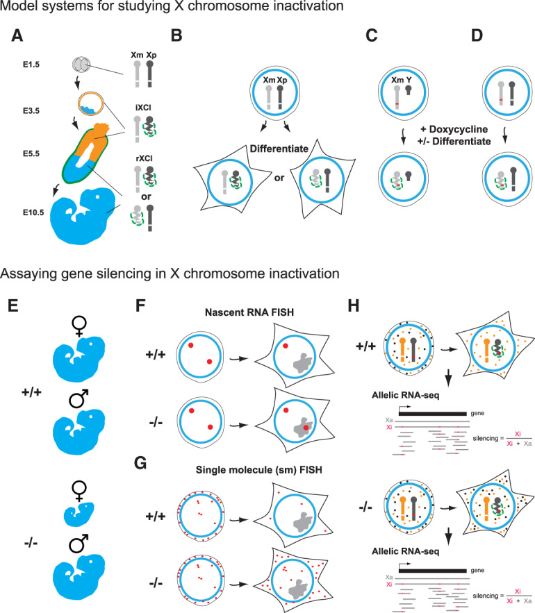
    1. Barros de Andrade ESL, Jonkers I, Syx L, Dunkel I, Chaumeil J, Picard C, Foret B, Chen CJ, Lis JT, Heard E, et al. 2019. Kinetics of Xist-induced gene silencing can be predicted from combinations of epigenetic and genomic features. Genome Res 29: 1087–1099. 10.1101/gr.245027.118 - DOI - PMC - PubMed

Publication types

MeSH terms

LinkOut - more resources